Structures and Functions in Living Organisms · 6 question types
Past paper frequency (2018 to 2024)
This topic accounts for approximately 15% of your exam marks.
The heart, blood vessels, and blood components are regularly tested, particularly structure-function links.
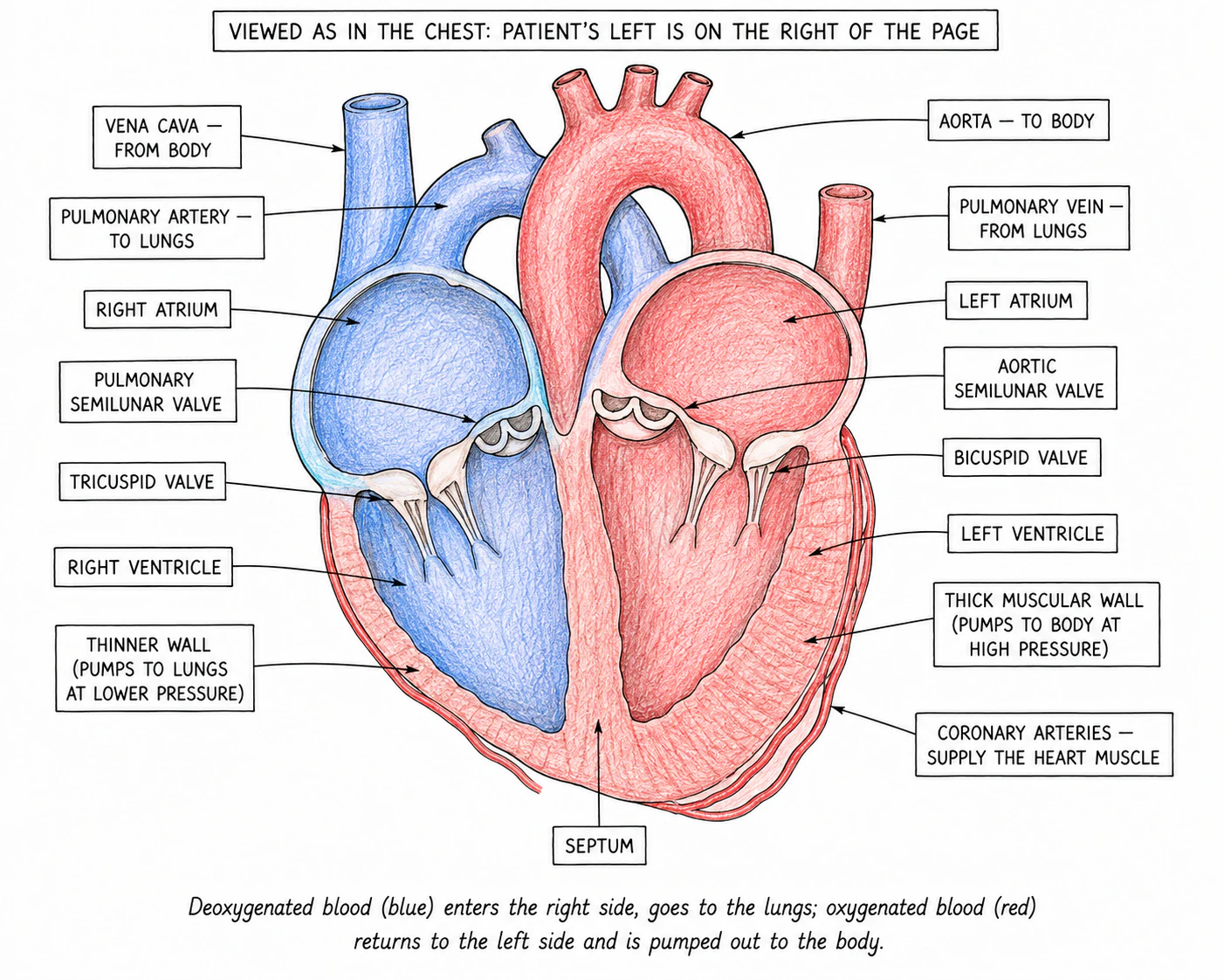
The heart is a muscular pump about the size of a closed fist, sitting just left of centre in the chest. It is really two pumps stuck together, working side by side: a right side that pumps deoxygenated blood to the lungs, and a left side that pumps oxygenated blood to the rest of the body.
Each side of the heart has two chambers:
| Chamber | What it does |
|---|---|
| Right atrium | Receives deoxygenated blood from the body via the vena cava |
| Right ventricle | Pumps that deoxygenated blood to the lungs via the pulmonary artery |
| Left atrium | Receives oxygenated blood from the lungs via the pulmonary vein |
| Left ventricle | Pumps that oxygenated blood to the whole body via the aorta |
The atria are the small upper chambers that receive blood from the veins. The ventricles are the larger lower chambers that pump blood out into the arteries.
The two sides are completely separated by a thick muscle wall called the septum. This separation keeps oxygenated and deoxygenated blood from mixing.
The left ventricle is built with a much chunkier muscle wall than its right counterpart. The reason is workload: the left ventricle has to drive blood at high pressure all the way around the whole body (the systemic circuit), while the right ventricle only sends blood a short way to the lungs (the pulmonary circuit). A thicker, stronger muscle generates the higher pressure.
The right ventricle deliberately pumps at lower pressure: the delicate capillaries in the alveoli of the lungs would be damaged by high-pressure blood.
Why the left ventricle wall is thicker
Explaining why the left ventricle wall is thicker comes up (3 marks), so you need to know it has more muscle to generate greater pressure, because it pumps blood all around the body (the right ventricle only pumps to the lungs). Say it generates pressure — "withstands high pressure" scores nothing.
The heart has four one-way valves that stop blood from flowing backwards:
| Valve | Where it is | What it does |
|---|---|---|
| Right atrioventricular valve (tricuspid) | Between right atrium and right ventricle | Stops blood flowing back from the right ventricle into the right atrium when the ventricle contracts |
| Left atrioventricular valve (bicuspid / mitral) | Between left atrium and left ventricle | Stops blood flowing back from the left ventricle into the left atrium |
| Semilunar valve (pulmonary) | At the start of the pulmonary artery |
| Stops blood flowing back from the pulmonary artery into the right ventricle |
| Semilunar valve (aortic) | At the start of the aorta | Stops blood flowing back from the aorta into the left ventricle |
Starting from where blood arrives back from the body:
In reality both atria contract together, then both ventricles contract together; the diagram describes one side at a time only for clarity.
The heart muscle itself needs a constant supply of oxygen and glucose for its own respiration. These are delivered by the coronary arteries, which branch off the aorta as soon as it leaves the heart and run across the outside of the heart muscle. If a coronary artery becomes blocked, the patch of heart muscle it feeds runs out of oxygen and dies, causing a heart attack (section 7).