Transport in Humans
Structures and Functions in Living Organisms · 6 question types
Humans use three kinds of blood vessel, each shaped for one job:
| Feature | Artery | Vein | Capillary |
|---|---|---|---|
| Direction of flow | Away from the heart | Towards the heart | Between artery and vein, through tissues |
| Blood pressure | High (the heart's pump has just pushed blood out) | Low (most of the pressure has been lost in the capillaries) | Low |
| Wall thickness | Thick, muscular, with elastic fibres | Thin | One cell thick |
| Lumen (the inner channel) | Narrow | Wide | Very narrow (only just wide enough for a single red blood cell) |
| Valves? | No (the semilunar valves at the start of the aorta and pulmonary artery belong to the heart, not to the arteries themselves) | Yes, frequent pocket-shaped valves | No |
| Blood content | Usually oxygenated (exception: pulmonary artery) | Usually deoxygenated (exception: pulmonary vein) | Both oxygenated and deoxygenated, depending on where in the body |
A handy memory trick: Arteries carry blood Away from the heart.
Why each vessel has the structure it does
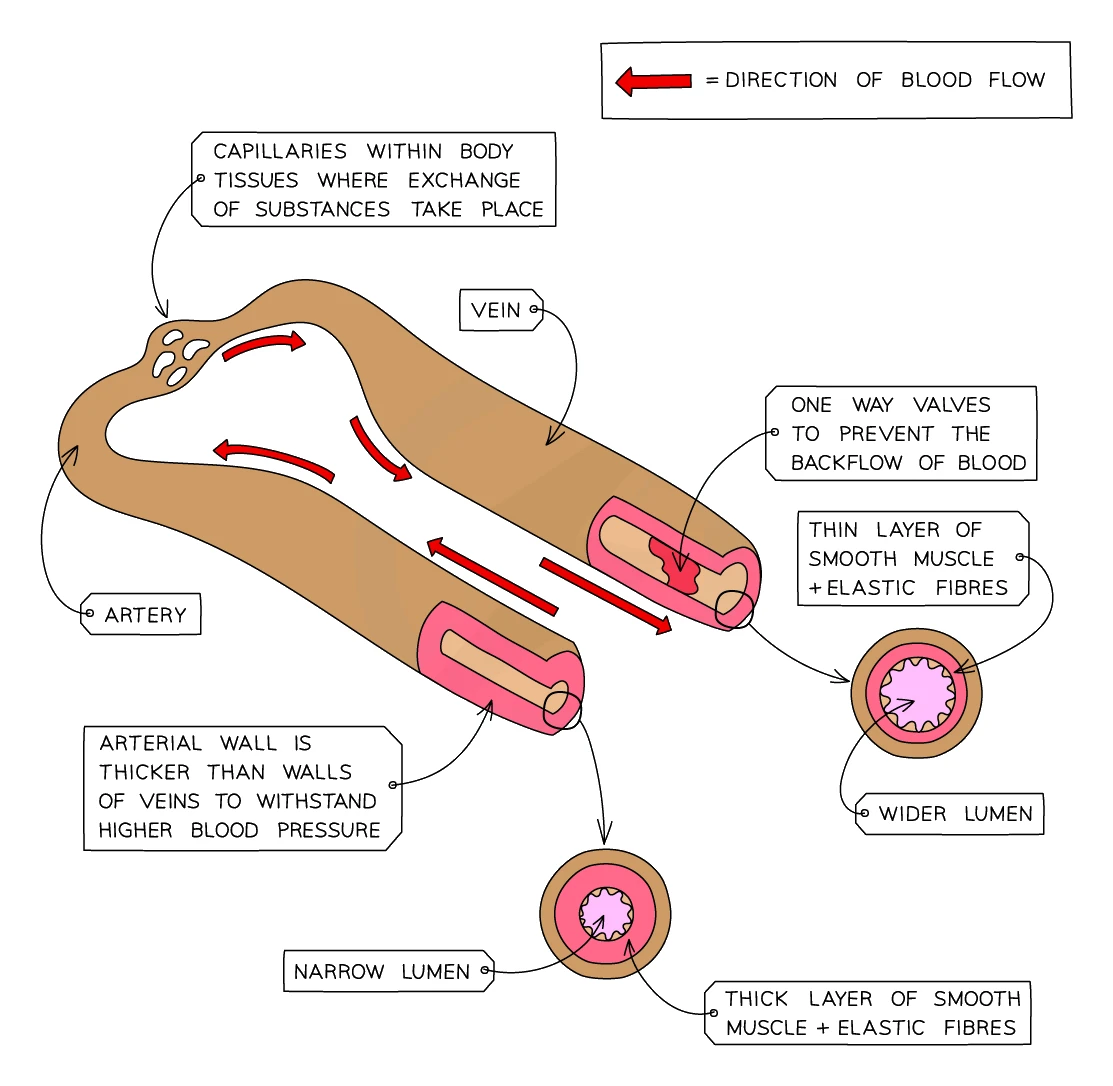
Arteries have to cope with the high pressure of blood freshly pumped from the heart:
- Thick muscular wall withstands the pressure without bursting
- Elastic fibres in the wall stretch when the heart beats and recoil between beats, evening out the surges of pressure
- Narrow lumen keeps the pressure high so blood is delivered quickly to distant tissues
Veins carry blood back to the heart under much lower pressure:
- Thinner wall is enough for the low pressure
- Wider lumen offers less resistance to the slow blood flow
- Valves stop the blood from flowing backwards. When you tense the muscles around a vein (e.g. by walking), the squeezing pushes blood forwards; the valves stop it sliding back when the muscle relaxes
- Many large veins run between skeletal muscles, so movement of the limbs helps pump the blood back to the heart
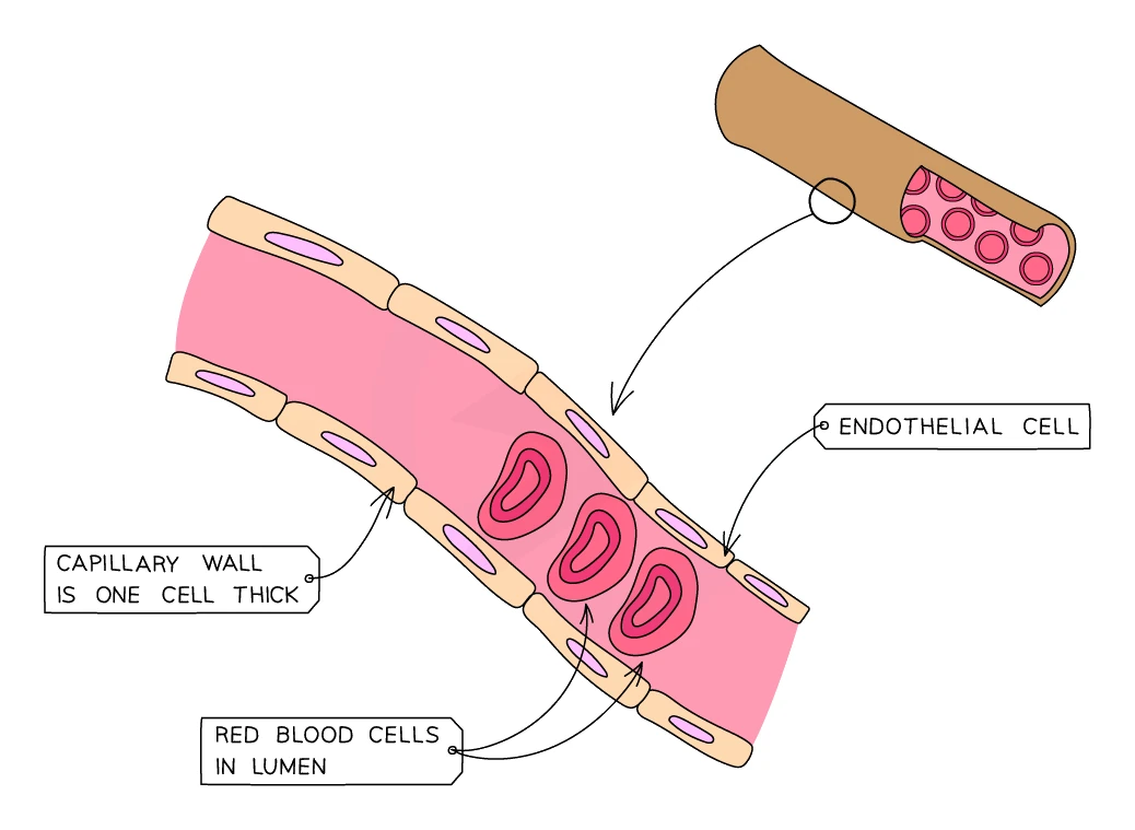
Capillaries are where the actual exchange between blood and tissues happens. They form a dense network branching out from arteries and joining back up into veins inside every tissue:
- One cell thick: the wall is a single layer of cells, so the diffusion distance for oxygen, glucose, CO₂ and urea is as short as possible
- Very narrow lumen forces red blood cells to pass through one at a time, slowing the flow and giving plenty of time for diffusion
- "Leaky" walls allow plasma to seep out into the surrounding tissue as tissue fluid, which bathes the cells and delivers their supplies. Most tissue fluid is reabsorbed; the rest is collected by a separate network of lymphatic vessels and returned to the bloodstream
Explaining differences between an artery and a vein
What comes up: "explain two differences between the structure of the wall of an artery and a vein" (4 marks, marked in pairs — each structural point earns 1 mark, each linked reason earns a second mark).
Write (four marks): Give two paired points. For example: (1) An artery has a thicker layer of elastic fibres (1) because the blood pressure is higher and the wall must recoil to smooth the flow (1). (2) An artery has a thicker layer of muscle (1) to control blood flow (1). Alternatively, replace either pair with: arteries have no valves (1) because blood pressure is sufficient to prevent backflow (1).
Watch out: simply writing "thick wall due to high pressure" gains only two marks if you do not separate the elastic and muscular layers. State each layer as a distinct point and give a separate reason for each to reach full marks.