Nutrition & Digestion
Structures and Functions in Living Organisms · 9 question types
Once digestion is complete, the small soluble products (glucose, amino acids, fatty acids and glycerol) need to be absorbed into the blood. This happens in the ileum, the long second part of the small intestine.
Adaptations of the small intestine for absorption
The ileum is brilliantly adapted to take in the maximum amount of dissolved food in the shortest time:
- Very long (around 6 metres in an adult): the food spends a long time in contact with the absorbing surface
- Inner surface folded into millions of tiny finger-like projections called villi: each villus is about 1 mm long
- The cells covering each villus have their surfaces folded into thousands of even tinier
- Combined, the villi and microvilli increase the inner surface area to roughly 250 m², about the size of a tennis court
Structure of a villus
Each villus has:
- A thin wall just one cell thick (a single layer of epithelial cells), so the diffusion distance for absorbed molecules is very short
- A dense network of blood capillaries running just inside the wall, which carries away the glucose and amino acids as soon as they are absorbed. This keeps the concentration gradient between the gut and the villus steep, maintaining fast absorption
- A (a small lymph vessel) running up the centre, which absorbs the fatty acids and glycerol and carries them into the lymphatic system
Most digested food is absorbed by diffusion (down its concentration gradient into the blood). When the gradient runs out, active transport is used to absorb the last molecules against the gradient.
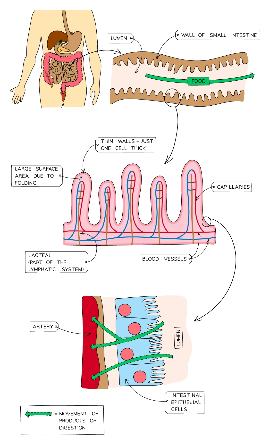
Adaptations of the small intestine for absorption
What comes up: "Explain how the structure of the small intestine is adapted for absorption" — typically a 4-mark question requiring structure + linked explanation for each point.
Write (four marks, each needs structure AND reason): (1) villi (finger-like projections) increase the surface area for absorption; (2) microvilli on the surface of villus cells further increase the surface area; (3) the wall is one cell thick, giving a short diffusion distance so molecules cross quickly; (4) a network of blood capillaries inside each villus carries absorbed glucose and amino acids away, maintaining a steep concentration gradient so diffusion continues; (5) a central lacteal absorbs fatty acids and glycerol into the lymphatic system.
Watch out: The mark scheme requires both the structural feature and the functional explanation for each mark — naming "villi" alone, without linking it to increased surface area, gains no credit. Similarly, stating "thin wall" must be paired with "short diffusion distance" or equivalent.