Exam Frequency Analysis
Past paper frequency (2018 to 2024)
This topic accounts for approximately 14% of your exam marks.
stable
High
Stable14%
Nervous system structure, reflex arcs, and hormones are all commonly examined.
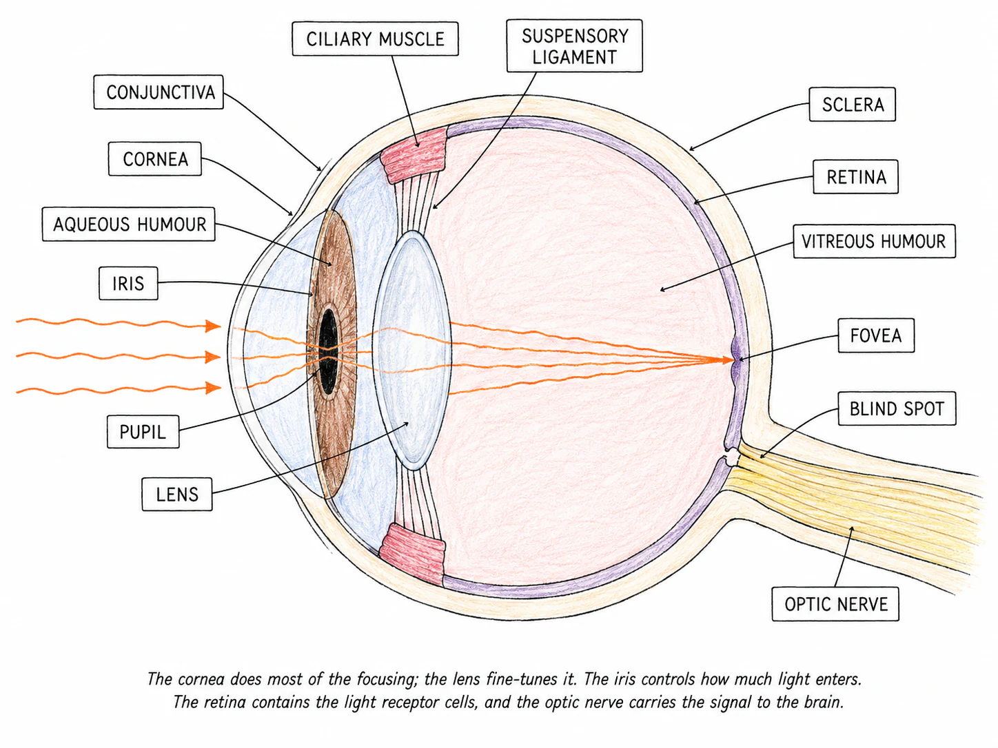
The eye is a sense organ that detects the stimulus of light. It contains light-sensitive receptor cells, plus a sophisticated lens system that focuses light onto those cells.
The main structures of the eye
| Structure | What it does |
|---|---|
| Cornea | A see-through, dome-shaped tissue at the very front of the eyeball. Refracts (bends) light as it enters, doing most of the focusing |
| Sclera | The tough white outer wall of the eyeball. Keeps the eye's shape; provides attachment for the muscles that move the eye |
| Conjunctiva | A thin transparent membrane covering the front of the eye; keeps it moist and protected |
| Iris | The coloured ring of muscle that controls how much light enters by changing the size of the pupil |
Accommodation: focusing on near and far objects
The eye uses two parts to focus light: the cornea does most of the bending, and the lens does the fine-tuning. The lens can change shape, which lets the eye focus on objects at different distances. This is called .
Looking at a near object (e.g. reading a book):
- The ciliary muscle contracts, so the ring of muscle becomes smaller in diameter
- This slackens the suspensory ligaments
- The lens, no longer pulled tight, becomes fatter and more rounded
- A fatter lens refracts (bends) the light more, focusing the light from the close object onto the retina
Looking at a far object (e.g. a tree on a distant hill):
- The ciliary muscle relaxes, so the ring of muscle becomes wider in diameter
- This pulls the suspensory ligaments tight
- The taut ligaments pull on the lens, making it thinner and flatter
- A thinner lens refracts the light less, which is enough for the parallel rays of light from the distant object to focus on the retina
A common mistake: people sometimes write that "the suspensory ligaments contract". They don't, because they are ligaments, not muscles. The right words are tighten or slacken.
| Near object | Distant object | |
|---|---|---|
| Ciliary muscle | Contracts | Relaxes |
| Suspensory ligaments | Slack | Taut |
| Lens shape | Fat / rounded | Thin / flat |
| Light refraction |
Exam tip
Accommodation for a near object
Describing how the eye focuses on a near object is a 4-marker, so you need to know: the ciliary muscles contract, the suspensory ligaments slacken, the lens becomes fatter / more convex, and it refracts light more. Never say the suspensory ligaments "contract" or "relax" — their tension is reduced; it's the ciliary muscle that contracts.
The pupil reflex: responding to light intensity
The amount of light reaching the retina is controlled by the size of the pupil. The iris changes the pupil size automatically through a reflex, protecting the retina from too much light and helping with vision in dim light.
The iris contains two sets of muscles, working as antagonistic pairs:
- Circular muscles run around the pupil. When they contract, the pupil constricts (narrows).
- Radial muscles run from the centre outwards like spokes on a wheel. When they contract, the pupil dilates (widens).
In bright light:
- Photoreceptors detect high light intensity
- The circular muscles contract, the radial muscles relax
- The pupil constricts (becomes smaller)
- Less light enters the eye, protecting the retina from damage
In dim light:
- Photoreceptors detect low light intensity