Coordination & Response
Structures and Functions in Living Organisms · 7 question types
A reflex action is a very fast, automatic, involuntary response to a stimulus. You do not have to think about it; by the time you become aware of the stimulus, your body has already responded. Examples include pulling your hand away from a sharp object, blinking when something flies near your eye, coughing when food goes the wrong way, and the pupil narrowing in bright light.
Reflex actions are useful because they are fast and automatic, which protects the body from damage in dangerous situations.
The reflex arc
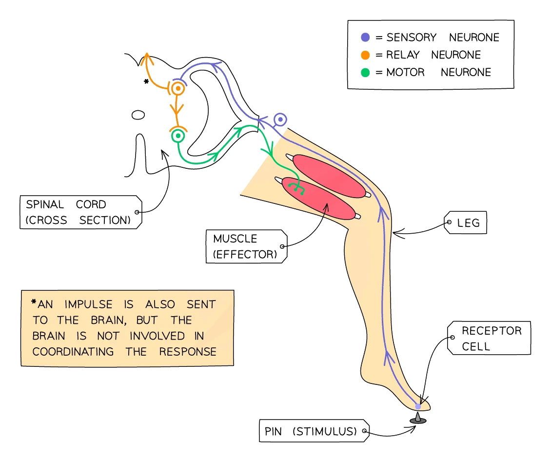
The neural pathway followed by a reflex action is called a . It bypasses the conscious part of the brain to save precious milliseconds. The arc usually goes through the spinal cord rather than the brain.
Tracing the reflex when a person steps on a sharp pin:
- Stimulus: the sharp point of the pin pressing on the foot
- Receptor: pain receptors in the skin of the foot detect the pressure and damage
- Sensory neurone: an electrical impulse travels from the receptor up the leg into the spinal cord
- Relay neurone: inside the spinal cord, the sensory neurone synapses with a short relay neurone, which immediately synapses with a motor neurone. This is the arc: the signal arcs across the spinal cord without going up to the brain
- Motor neurone: an impulse travels back down the leg to the muscles
- Effector: the muscles in the leg contract
- Response: the foot is pulled up and away from the pin
The whole thing takes a fraction of a second. A separate, slower signal also goes up to the brain so that you become aware of the pain shortly after the foot has moved.
The reflex arc: stimulus → receptor → sensory neurone → relay neurone (in spinal cord) → motor neurone → → response
Describing the pathway of a reflex arc
What comes up: "Describe the pathway of a nerve impulse in the reflex arc" (4 marks) or "Name the structures involved in the withdrawal reflex in order."
Write (four marks): (1) A receptor detects the stimulus and generates an impulse. (2) The impulse travels along the sensory neurone to the CNS / spinal cord. (3) It passes to a relay neurone (located inside the spinal cord), via a synapse. (4) The impulse travels along a motor neurone to the effector (muscle or gland), which carries out the response.
Watch out: The relay neurone is specifically in the CNS (spinal cord) — do not place it outside the CNS. The mark scheme accepts "neurone" without qualification for one mark only; name each neurone type to score all marks.