1. Diffusion
What diffusion is
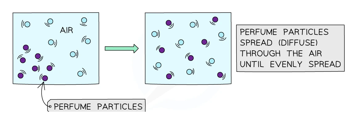
Diffusion is the net movement of particles from a region of higher concentration to a region of lower concentration, down a
Several things to notice about the definition:
- Net movement, not just movement. Individual particles bounce around randomly in all directions. The reason a substance appears to spread out is that, on average, more particles happen to move from the crowded side to the empty side than the other way around.
- Down a concentration gradient, which means from a high concentration to a low concentration. Diffusion never moves particles "uphill" against the gradient.
- It is a passive process, so it does not need any energy from the cell. The particles' own kinetic energy does all the work.
- Diffusion stops being net once the concentrations on the two sides are equal. The particles keep bouncing around after that, but with no net flow.
Why it happens
Every particle in a fluid is in constant, random motion. The more crowded a region is, the more particles in it bouncing around per second, and the more particles per second that happen to bump across into the neighbouring (less crowded) region. The reverse traffic is smaller because fewer particles live in the lower-concentration region in the first place. Over time, the imbalance evens out.
Diffusion across cell membranes
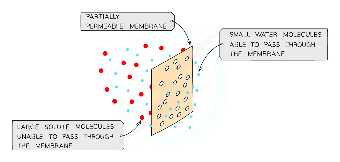
The cell membrane is partially permeable (sometimes called selectively permeable). It lets some substances cross freely but blocks others:
- Small, uncharged molecules like oxygen, carbon dioxide, water, urea and small lipid-soluble molecules diffuse across the membrane with no help
- Large molecules like starch, glucose and proteins cannot diffuse across on their own
- Charged ions also struggle to cross the lipid membrane without help
Important examples of diffusion in living organisms
| Where | What diffuses | Why a gradient exists |
|---|---|---|
| Alveoli in the lungs | Oxygen in to the blood; carbon dioxide out | Air in alveoli is rich in O₂ and poor in CO₂; blood arriving in the capillaries is the opposite |
| Photosynthesising leaf | Carbon dioxide diffuses into the leaf; oxygen diffuses out | Chloroplasts use up CO₂ and produce O₂, keeping the gradient steep |
| Liver to bloodstream | Urea leaves the liver cells and enters the surrounding blood by diffusion | The liver produces urea continuously, so its concentration inside the cells stays high while the blood washes it away |
| Plant root | Water and dissolved gases | Wet soil has higher concentrations than the inside of the root |
2. Osmosis
What osmosis is
Osmosis is the net movement of water molecules from a region of higher water concentration (a dilute solution) to a region of lower water concentration (a concentrated solution), across a
Osmosis is really just a special case of diffusion: it is the diffusion of water through a partially permeable membrane. The membrane has to be partially permeable because that is the only way to keep two solutions with different concentrations of solute separate; if everything could cross freely, the solutes would simply diffuse out and equalise.
Water potential: a more accurate way to talk about it
Some textbooks use the term instead of water concentration. The idea is the same:
- Pure water has the highest water potential (the highest "willingness" of water to leave)
- The more solute dissolved in a solution (sugar, salt, etc.), the lower its water potential
- Water always moves from higher water potential to lower water potential by osmosis
Both ways of describing osmosis are accepted in exam answers. Using water potential is slightly more accurate.
Exam tip
Give both halves of the osmosis definition
A definition of osmosis is worth two marks: one for the movement of water through a partially permeable membrane ("semi-permeable" and "selectively permeable" are accepted) and one for the direction, from a dilute solution to a concentrated one or from higher to lower water potential. "From a high concentration of water to a low concentration of water" is accepted for the direction, and one sentence covering both the membrane and the direction earns both marks. Leave out the membrane, or reverse the direction, and that half's mark is gone.
Osmosis in animal cells
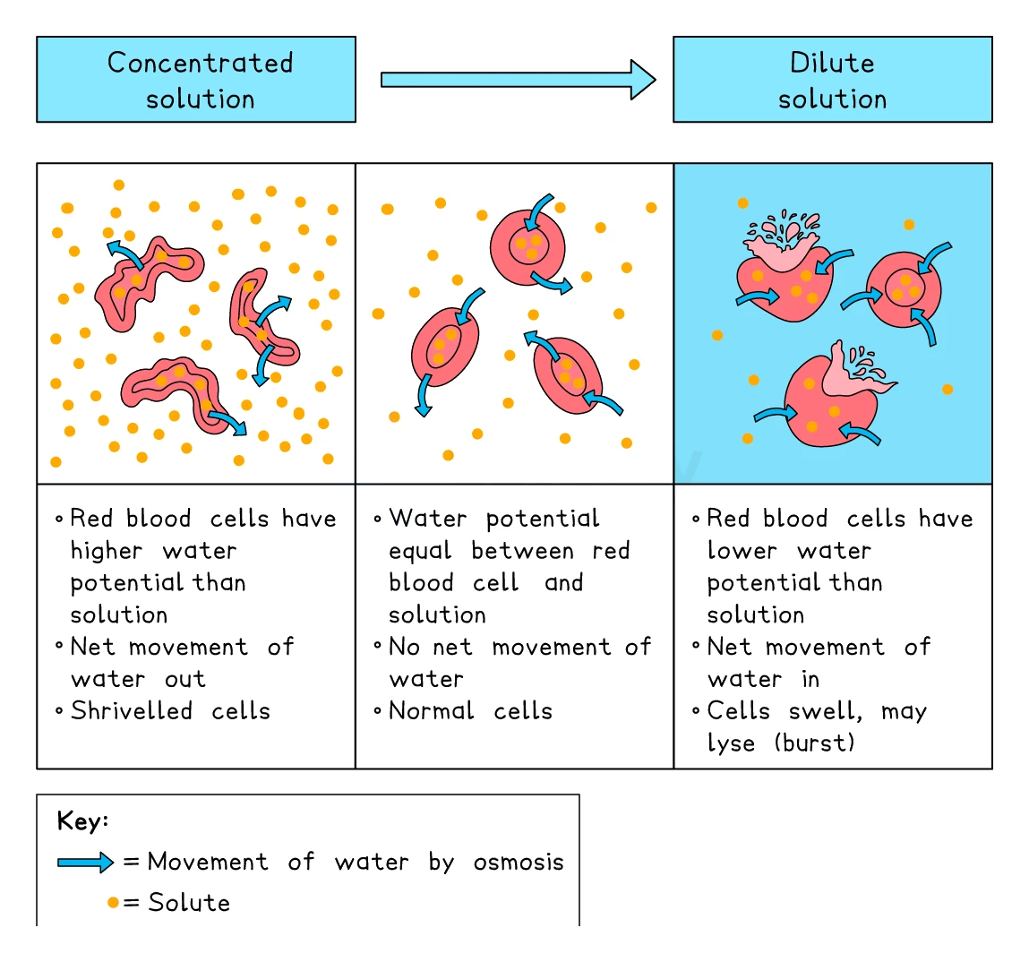
Animal cells have no cell wall, so what happens to them in different solutions is dramatic:
| External solution | What happens to the cell | Cell state |
|---|---|---|
| Distilled water (higher water concentration outside) | Water moves in by osmosis. The cell swells and eventually bursts because there is no wall to resist | Lysed (or haemolysed for red blood cells) |
| Solution with the same concentration as the cytoplasm | No net movement of water | Normal shape |
| Concentrated sugar or salt solution (lower water concentration outside) | Water moves out by osmosis. The cell shrivels | Crenated |
This is why doctors are careful about what solutions they use for intravenous drips: pure water would burst red blood cells, and very concentrated solutions would shrivel them.
Common exam question
Explaining why red blood cells burst in water
Question: Explain what happens to red blood cells in distilled water, or why cells are kept in salt solution rather than pure water (2–3 marks).
Asked in 5 of the 23 papers. The marks form a chain: water enters the cell by osmosis, from the higher water potential outside to the lower water potential inside (or dilute to concentrated), and with no cell wall the cell cannot stop expanding, so it bursts. Osmosis, the direction and the bursting are separate marks. Burst cells vanish, and the cloudy suspension becomes a clear red solution as haemoglobin dissolves.
Stored in a solution matching the cytoplasm, cells take up no net water by osmosis and do not burst. In concentrated salt solution, water leaves by osmosis and the cells shrivel (crenate); "plasmolysed" is rejected and "flaccid" ignored. When a question contrasts them with bacteria, one mark is for which cell has a wall; saying water does not enter the walled cell is ignored.
Osmosis in plant cells
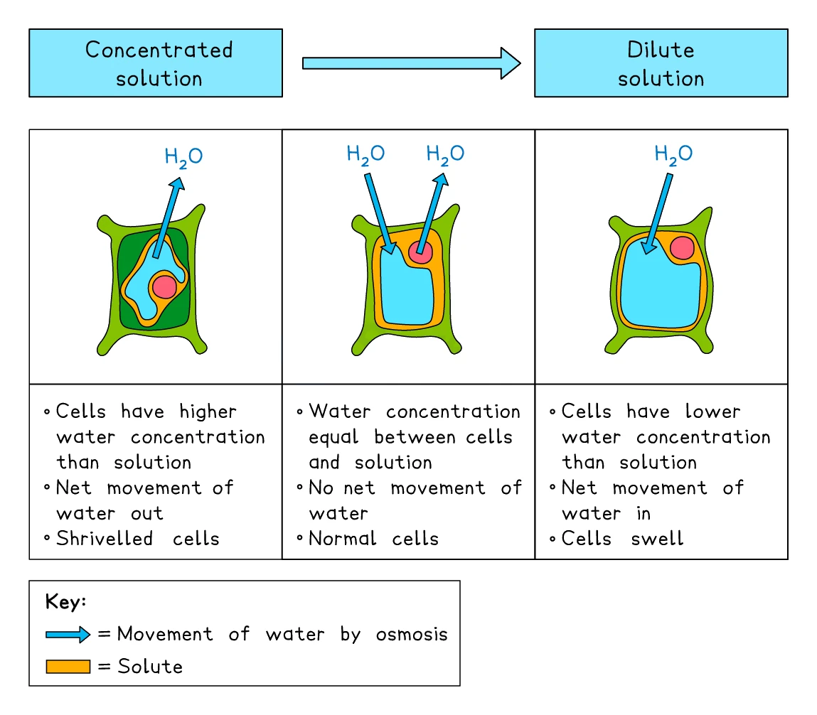
Plant cells have a tough cellulose cell wall that prevents bursting, so the same situations produce different results:
| External solution | What happens to the cell | Cell state |
|---|---|---|
| Distilled water | Water moves in. The vacuole fills up and pushes the cytoplasm hard against the cell wall. The cell becomes firm but does not burst because the wall holds | Turgid (the desired state; keeps plants upright) |
| Solution at the same concentration as the cytoplasm | No net movement | Normal but limp |
| Concentrated solution | Water moves out. The vacuole shrinks; the cytoplasm pulls away from the wall. The cell becomes limp | Flaccid, and at extremes plasmolysed (membrane separated from wall) |
A wilting houseplant is showing flaccid cells. Watering it brings the cells back up to turgid and the plant stands up again.
Common exam question
Explaining plant cells in water and in a strong solution
Question: Explain the difference in appearance between a plant cell in distilled water and one in a concentrated salt solution, or the change in onion cells after an hour in sucrose solution (3–4 marks).
Asked in 2 of the 23 papers. In distilled water, water enters the cell, by osmosis, from the dilute solution to the more concentrated cell contents (or from higher to lower water potential), so the cytoplasm is pushed against the cell wall and the cell is turgid. In the concentrated solution the same points run the other way: water leaves by osmosis, down the water potential gradient, the membrane pulls away from the wall and the cell is flaccid or plasmolysed.
"By osmosis" and the direction are separate marks, so name the process and give the direction every time. "The cell shrinks" on its own is ignored; say what the cytoplasm or the membrane does.
3. Active transport
What active transport is
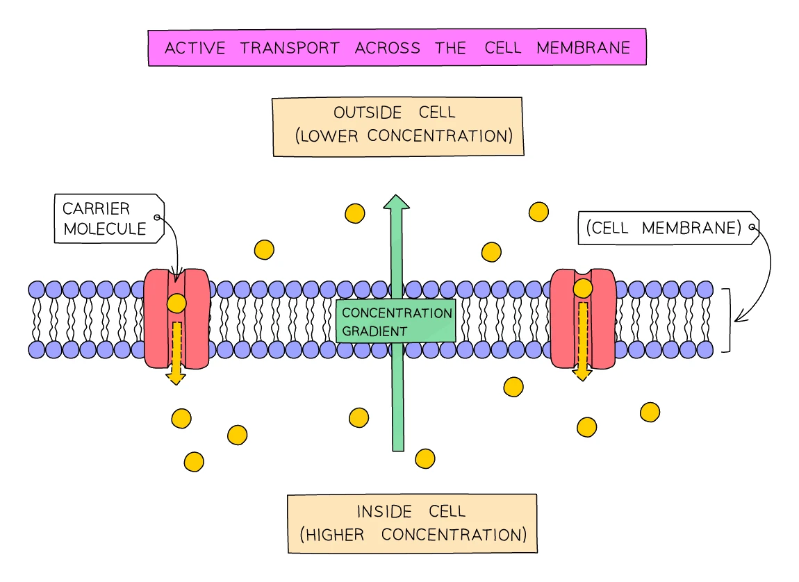
Active transport is the movement of particles across a cell membrane from a region of lower concentration to a region of higher concentration, against the concentration gradient, using energy released by respiration
Two things make active transport different from diffusion and osmosis:
- The direction is against the . Particles are being moved "uphill" from a place where they are scarce to a place where they are already concentrated.
- This needs energy from respiration (specifically, energy carried by ATP produced in the mitochondria). Cells that do a lot of active transport (root hair cells, gut lining cells, kidney tubule cells) are packed with mitochondria.
How it works at the molecular level
Active transport relies on protein pumps embedded in the cell membrane. Each pump:
- Binds the specific particle it transports on one side of the membrane
- Uses energy from ATP to change shape
- Releases the particle on the other side of the membrane (the high-concentration side)
A single pump can move thousands of particles per second when supplied with enough ATP.
Important examples of active transport
- Root hair cells in plants absorb mineral ions (nitrate, magnesium, potassium, etc.) from the soil. Soil water typically has very low ion concentrations, while the root cells have much higher concentrations inside. The plant cannot rely on diffusion; it has to pump the ions in against the gradient.
- Cells lining the small intestine absorb glucose and amino acids from digested food. By the end of digestion, the gut contents have a lower concentration of these molecules than the blood, but the body still needs to take in every last molecule. Active transport pumps them in.
- Kidney tubule cells reabsorb glucose from the filtrate back into the blood by active transport, even though the glucose in the filtrate is at a lower concentration than in the blood.
Common exam question
Why waterlogged roots take up fewer mineral ions
Question: Explain how overwatering, which fills the air spaces in the soil, can stop a plant growing properly or taking up mineral ions (2–3 marks).
Asked in 2 of the 23 papers. The marks are a chain: waterlogged soil has no oxygen around the roots, so the root cells cannot respire aerobically and release less energy as ATP, so they cannot carry out active transport, so they absorb fewer mineral ions. Each link can be a mark, so write the whole chain rather than jumping from "no oxygen" to "fewer minerals".
Wherever active transport is the credited process (mineral ions into root hair cells, glucose reabsorbed in the kidney), add that it uses energy from ATP and moves the substance against the concentration gradient: in the kidney schemes each of those earns a mark of its own in one paper or another. Active transport uses ATP but does not make it; respiration does, as one multiple-choice question has tested.
Side-by-side comparison
| Diffusion | Osmosis | Active transport | |
|---|---|---|---|
| What moves | Any particles (gases, small molecules) | Water only | Specific particles (often ions, glucose, amino acids) |
| Direction | High to low concentration | High to low water concentration (high to low water potential) | Low to high (against the gradient) |
| Needs a membrane? | No (happens anywhere) | Yes (partially permeable) | Yes (carries the protein pumps) |
| Energy required? | No (passive) | No (passive) | Yes (from respiration, via ATP) |
| Speed | Slows as gradient narrows | Slows as gradient narrows | Steady, set by the rate the pumps work |
| Example | O₂ into the blood at the alveoli | Water into root cells from wet soil | Mineral ions into root hair cells from soil |
4. Factors that affect the rate of diffusion
Four factors set how fast diffusion happens. Each one shows up in real exchange surfaces in the body.
Concentration gradient
The steeper the difference in concentration between the two sides, the faster the rate of diffusion. If you double the concentration on the high side (and keep the other side the same), you roughly double the net rate of particles crossing.
Real-world example: blood flow through the alveoli is continuous, so blood low in oxygen is always arriving. This keeps the oxygen gradient between the alveolar air and the blood steep, maintaining a fast rate of oxygen diffusion into the blood.
Exam tip
Say what keeps the concentration gradient steep
In any question about an exchange surface (alveoli, villi, a dialysis machine, a leaf losing water) there is usually a mark for the gradient, and it is earned by saying what maintains it: the blood supply carrying absorbed substances away, dialysis fluid being pumped past the membrane, or wind moving humid air off the leaf. Schemes mark these points in pairs, a structure with its function, and one refuses the function mark without the structure, so put the blood supply and the steep gradient it maintains in the same sentence.
Temperature
When a substance is hotter, its particles carry more kinetic energy and zip around at higher speeds. Faster particles produce more collisions per second against any boundary, which means a faster rate of diffusion.
This is why a teaspoon of sugar dissolves much faster in hot tea than in cold water.
Surface area
The larger the surface area of the membrane through which particles can diffuse, the more particles can cross per second.
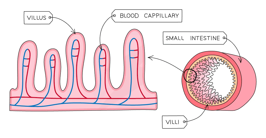
Real-world example: the human small intestine has its inner surface folded into villi, and each villus is covered with microvilli, giving a total surface area of around 250 m² (the size of a tennis court) packed inside the abdomen. This enormous surface area maximises the rate at which digested food can be absorbed.
Diffusion distance
The thinner the layer the particles have to cross, the faster they finish the journey. Rate of diffusion is roughly inversely proportional to the thickness of the barrier.
Real-world example: the wall of an alveolus is just one cell thick, and the wall of the surrounding capillary is also just one cell thick. The total gas-exchange distance is therefore only about 0.5 μm, which is short enough for gases to cross in a fraction of a second.
Surface area to volume ratio
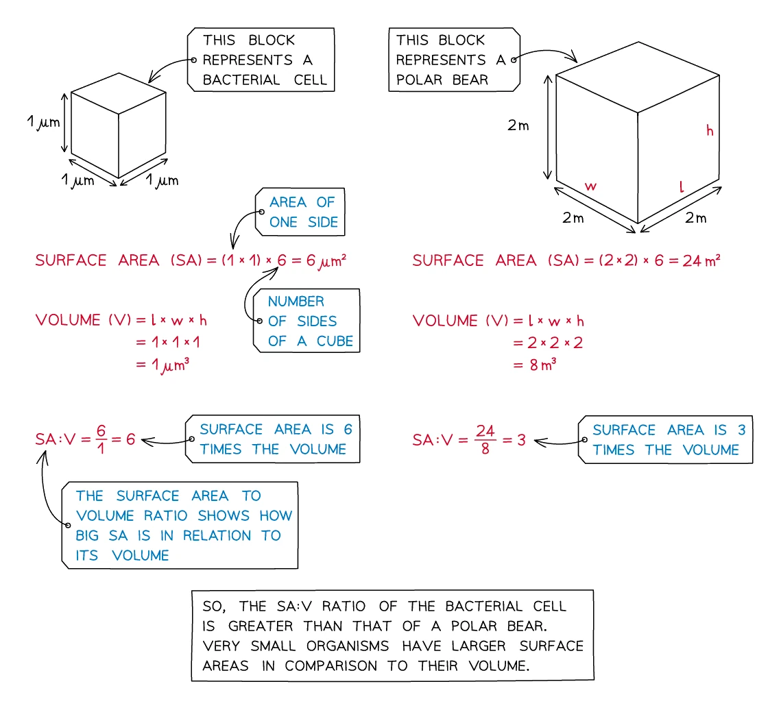
When considering whole cells or whole organisms, all four factors combine into one big principle: surface area to volume ratio (SA:V). As an object gets larger:
- Its volume grows with the cube of its linear size
- Its surface area grows with the square of its linear size
- So its SA:V ratio falls as it gets bigger
A larger object has less surface per unit of volume to take in supplies and get rid of waste. Single-celled organisms (bacteria, amoebas) can live on diffusion alone because they are so small that their SA:V ratio is huge. Larger organisms need specialised exchange surfaces (alveoli, villi, root hairs, gills) to overcome the falling SA:V ratio.
Worked example
Surface area to volume ratio of a cube-shaped cell
A cell is roughly a cube with sides of 0.04 mm. Calculate its surface area to volume ratio, giving your answer in the form n : 1.
Solution:
- Surface area = 6 × 0.04 × 0.04 = 0.0096 mm²
- Volume = 0.04 × 0.04 × 0.04 = 0.000064 mm³
- Ratio = 0.0096 ÷ 0.000064 = 150, so the surface area to volume ratio is 150 : 1
Keep every decimal place until the division, or write the two values in standard form (9.6 × 10⁻³ mm² and 6.4 × 10⁻⁵ mm³). The surface area and the volume each earn a mark on their own, a correct ratio with no working earns all three, and an answer rounded to the same number of significant figures as the data is accepted.
5. Core practical: investigating diffusion using agar cubes
Aim
To investigate how surface area to volume ratio affects the rate of diffusion.
Principle
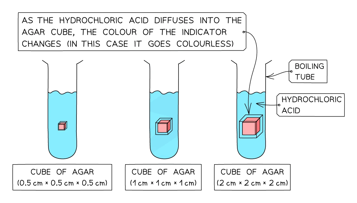
Cubes of agar jelly are coloured pink using sodium hydroxide (an alkali) and phenolphthalein (an indicator that is pink in alkaline conditions and colourless below pH 8.3). When placed into dilute hydrochloric acid, the acid diffuses inwards through the agar and neutralises the alkali. As it does, the indicator turns colourless. The time it takes for the cube to become completely colourless is a measure of how long it took the acid to diffuse to the centre.
Cubes of different sizes have different surface area to volume ratios, so they decolourise at different rates.
Apparatus
- Block of pink agar (already made up with sodium hydroxide and phenolphthalein)
- White tile (to provide a clean cutting surface and contrast)
- Sharp scalpel
- Ruler
- Forceps
- Dilute hydrochloric acid (around 1 mol/dm³)
- Boiling tubes, one per cube size
- Stopwatch
Method
- On the white tile, use the scalpel and ruler to cut three cubes of pink agar of different side lengths. A useful set of sizes is 1 cm, 2 cm and 3 cm.
- For each cube, calculate the surface area, the volume and the SA:V ratio. Record these in a table.
- Pour the same volume of dilute hydrochloric acid into each boiling tube (around 30 ml).
- Using forceps, place the smallest cube into one boiling tube and start the stopwatch at the same time.
- Watch the cube. Stop the stopwatch when the cube has turned completely colourless. Record the time.
- Repeat steps 4 and 5 for the medium and large cubes, using a fresh tube of acid each time.
- Repeat the whole experiment at least twice more so a mean time can be calculated for each cube size.
Variables
- Independent variable: cube size (SA:V ratio)
- Dependent variable: time taken for the cube to turn colourless
- Control variables: acid concentration, acid volume, temperature of the acid, type of indicator, shape of the cubes (all cubes), how thoroughly each cube is submerged
Sample results and analysis
Typical results would show:
| Cube side length (cm) | Surface area (cm²) | Volume (cm³) | SA:V ratio | Mean time to turn colourless (s) |
|---|---|---|---|---|
| 1 | 6 | 1 | 6:1 | shortest (e.g. 40 s) |
| 2 | 24 | 8 | 3:1 | medium (e.g. 110 s) |
| 3 | 54 | 27 | 2:1 | longest (e.g. 220 s) |
The smaller the cube, the higher its SA:V ratio, the shorter the distance from the surface to the centre, and so the faster the acid diffuses through to neutralise all the alkali.
Limitations
- It is hard to judge the exact moment a cube is "fully colourless", which introduces human error in timing. A measurable alternative is to leave each cube in acid for a fixed time (say 60 seconds), then cut it open and measure how far in (in mm) the pink colour has disappeared.
- It is hard to cut perfectly equal-sided cubes with a scalpel. Small differences in side length change the SA:V ratio noticeably.
- The cubes should be made of the same batch of agar so that the porosity is identical.
6. Core practical: investigating osmosis using potato cylinders
Aim
To investigate how the concentration of an external sucrose solution affects osmosis in plant cells.
Principle
Cylinders of fresh potato contain plant cells with cytoplasm at some particular water concentration. If a cylinder is placed into a solution of higher water concentration (dilute sucrose, or distilled water), water moves into the potato by osmosis, and the cylinder gains mass. If it is placed in a solution of lower water concentration (concentrated sucrose), water moves out, and the cylinder loses mass. Somewhere in the middle there is a sucrose concentration that exactly matches the water concentration inside the potato cells, and the cylinder shows no change in mass.
Apparatus
- A fresh potato
- Cork borer (to cut equal-diameter cylinders)
- Scalpel and ruler
- Five or six sucrose solutions of known concentration (typically 0.0, 0.2, 0.4, 0.6, 0.8 and 1.0 mol/dm³)
- Test tubes (one per solution) and a rack
- Mass balance, accurate to at least 0.01 g
- Paper towels
- Forceps
Method
- Use the cork borer to cut 6 equal-diameter cylinders of potato from the same potato.
- Use the scalpel and ruler to trim each cylinder to the same length (around 3 cm works well).
- Blot each cylinder dry on a paper towel.
- Weigh each cylinder on the balance and record its initial mass.
- Place one cylinder into each test tube containing a different sucrose concentration. Note carefully which cylinder is in which tube.
- Leave the tubes for at least an hour (longer is better; 24 hours gives the cleanest results).
- Remove each cylinder, blot it dry with a paper towel, and reweigh it.
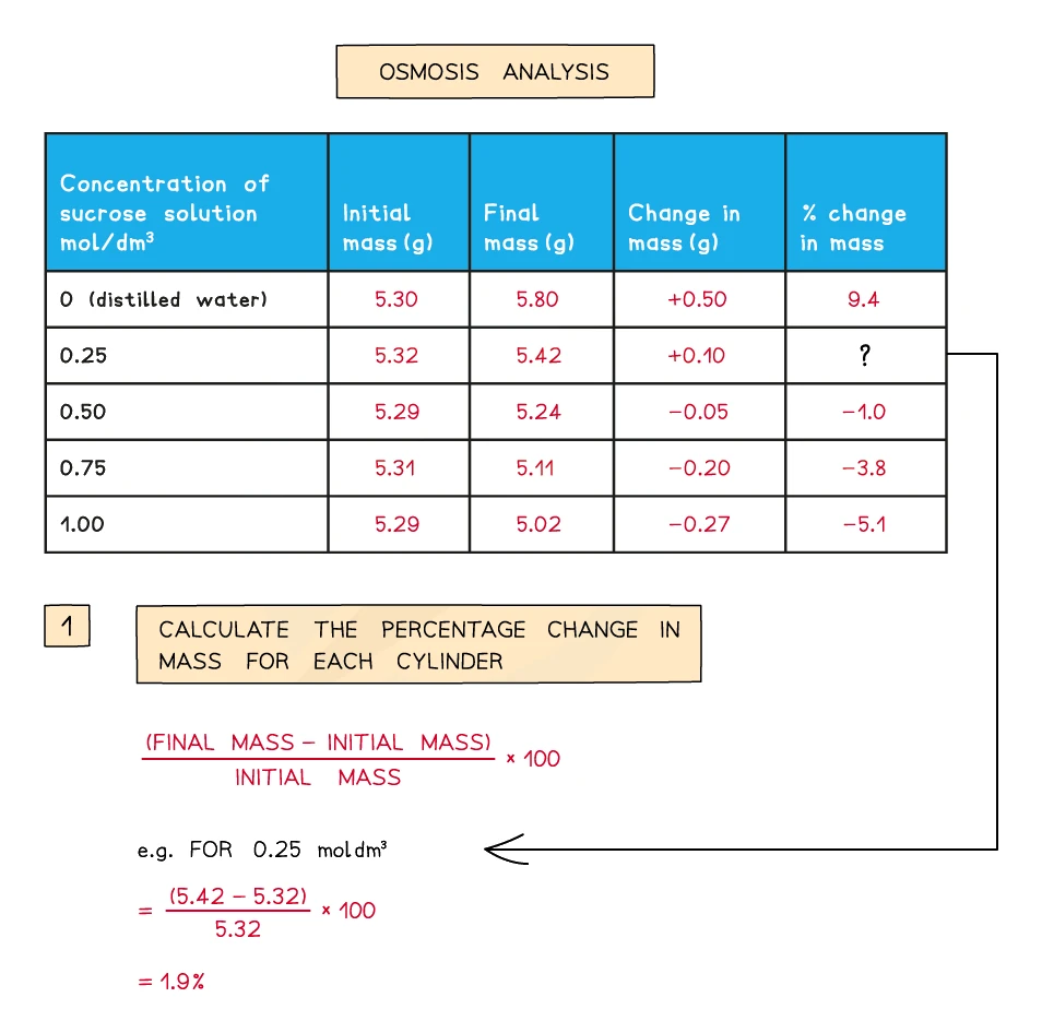
- Calculate the percentage change in mass for each cylinder using:
percentage change in mass = ((final mass − initial mass) ÷ initial mass) × 100
Variables
- Independent variable: sucrose concentration (mol/dm³)
- Dependent variable: percentage change in mass of the potato cylinder
- Control variables: type of potato, age of potato, length and diameter of cylinder, volume of solution in each tube, time left in solution, temperature, how each cylinder is blotted before weighing
Exam tip
Method questions on the potato practical
The independent variable is what is in each tube: the solution, or its concentration. For another variable to control, name one the method has not already fixed, such as temperature or type of potato; time, volume of solution and mass of potato are ignored, since the method already fixes or measures them. Drying the cylinders before weighing removes surface liquid that would add to the mass, so the masses can be compared fairly ("to remove water" is ignored, as are "reliable" and "accurate"). A larger surface area means more contact between potato and solution, so osmosis is faster.
Plotting and using the results
Plot percentage change in mass on the y-axis against sucrose concentration on the x-axis. The result is a straight-ish line falling from positive values on the left (low sucrose, water gained) through zero (no change) and on to negative values on the right (high sucrose, water lost).
The point where the line crosses the x-axis (zero change in mass) gives the sucrose concentration that matches the water concentration inside the potato cells. This is a clever way of measuring something you cannot weigh directly.
Common exam question
Explaining the change in mass of the potato cylinders
Question: Explain the changes in mass of potato cylinders left in distilled water, in air and in sucrose solutions of different concentrations (3–5 marks).
Asked in 2 of the 23 papers. Pair each result with its cause: the cylinder in distilled water or dilute sucrose gains mass because water enters it, the one in concentrated sucrose loses mass because water leaves, the movement is by osmosis, and it runs from higher to lower water potential (from the dilute to the concentrated solution). Each of those is a separate mark, so "osmosis" without the direction, or the direction without the word osmosis, leaves a mark behind.
Where one concentration gives no change in mass, say that there is no net movement of water because that solution has the same water potential as the potato: each half is a credited point. A cylinder left in air changes little because no osmosis happens; any small loss is evaporation.
Why use percentage change rather than absolute change?
Different cylinders start at slightly different masses no matter how carefully you cut them. Using percentage change cancels out those starting differences and lets you compare cylinders fairly.
Worked example
Calculating percentage change in mass of a potato cylinder
A potato cylinder has an initial mass of 5.0 g. After sitting in a concentrated sucrose solution for one hour, its final mass is 4.4 g. Calculate the percentage change in mass.
Solution:
- Change in mass = final mass − initial mass = 4.4 − 5.0 = −0.6 g
- Percentage change = (change in mass ÷ initial mass) × 100
- = (−0.6 ÷ 5.0) × 100 = −12%
The negative sign shows the cylinder lost mass: water moved out by osmosis into the more concentrated sucrose solution, and a positive answer would mean water moved in. Write the change in mass as a line of its own, because it earns a mark even if the division goes wrong, and a correct percentage with no working earns both marks.
Limitations
- Cylinders may differ slightly in dimensions, which affects how much water can be exchanged. Solution: cut several cylinders for each sucrose concentration and take a mean.
- Blotting before weighing has to be consistent. If one cylinder is more thoroughly dried than another, the mass reading is misleading.
- The potato has to be the same age and freshness throughout. Older potatoes have already lost water and behave differently.