1. Cells, organelles and the building blocks of life
Cells are the basic unit of life
All living organisms are built from one or more . A single-celled organism such as an amoeba is one cell on its own. A multicellular organism such as a human is built from trillions of cells working together.
Inside every cell is a jelly-like substance called the , surrounded by a thin that separates the cell from the outside world. Eukaryotic cells contain small sub-cellular structures called suspended in the cytoplasm; each organelle does a specific job.
The five levels of organisation
In a complex multicellular organism, structure is built up in five tidy levels:
| Level | What it is | Example |
|---|---|---|
| Organelle | A specialised sub-cellular structure inside a cell | Mitochondrion (site of aerobic respiration) |
| Cell | The basic structural and functional unit of life | Red blood cell |
| Tissue | A group of similar cells working together to perform one function | Muscle tissue, xylem tissue |
| Organ | Different tissues working together to perform a particular function | Heart, leaf, kidney |
| Organ system | A group of organs working together to perform a major function | Circulatory system, digestive system |
A handy way to remember the order: organelles → cells → → → .
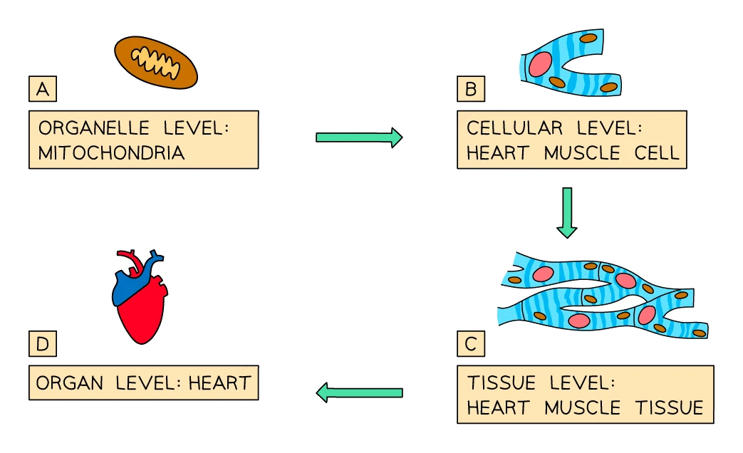
2. Organelles and their functions
The main organelles found in eukaryotic cells, with what each one actually does:
| Organelle | Function |
|---|---|
| Nucleus | Contains the cell's genetic material as chromosomes. Controls all of the cell's activities by switching genes on and off |
| Cytoplasm | A jelly-like fluid that fills the cell. The site of most chemical reactions, including the first stage of respiration. Contains water with many dissolved substances |
| Cell membrane | A thin layer of lipids and proteins. Controls what enters and leaves the cell. Partially permeable |
| Mitochondria | The site of aerobic respiration. Cells with high energy demand (muscle, liver, sperm) contain many mitochondria |
| Ribosomes | The site of protein synthesis. Tiny dots scattered through the cytoplasm or attached to membranes |
| Cell wall (plants only) | Made of cellulose. Gives the cell a fixed shape and stops it bursting when full of water |
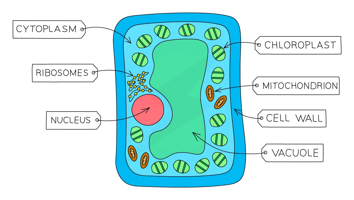
| Chloroplasts (plants only) | The site of photosynthesis. Contain the green pigment chlorophyll that absorbs light energy. Found in leaf and stem cells, not in roots |
| Permanent vacuole (plants only) | A large fluid-filled sac containing cell sap (water + dissolved sugars and salts). Its outward pressure squashes the cytoplasm up against the inside of the cell wall, leaving the cell firm and turgid, which helps the plant stand upright |
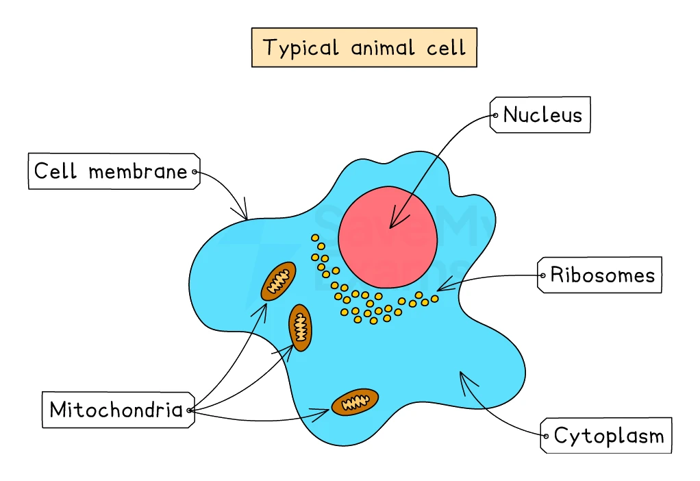
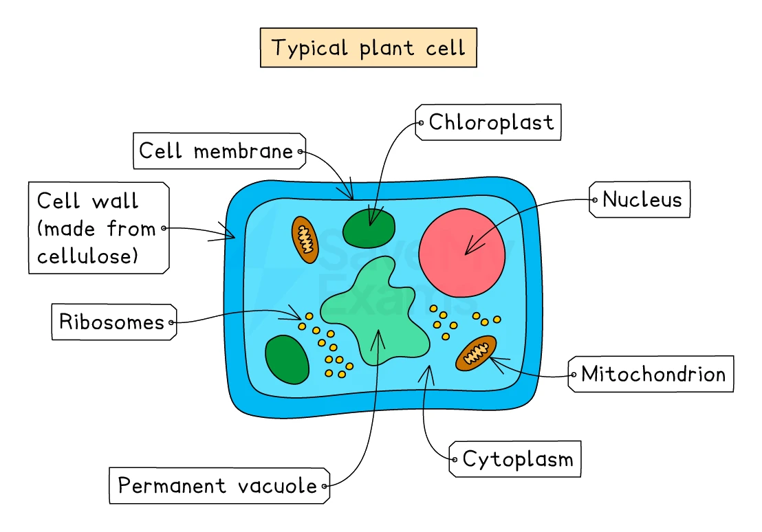
A quick note on terminology: mitochondrion is the singular (one organelle), mitochondria is the plural. The same pattern applies to / nuclei. Chloroplast / chloroplasts is different: it just takes a regular English plural by adding -s.
Common exam question
Identifying a labelled organelle or giving its function
Question: On a labelled drawing of a plant, animal or bacterial cell, say which letter is the nucleus, vacuole or cell wall, name the structure that does a given job, or describe what one organelle does (1–2 marks).
Asked in 8 of the 23 papers, usually as multiple-choice parts. Read the drawing by shape: the large clear space in a plant cell is the vacuole (the scheme credits "cell sap" for it too), the small dense body is the nucleus, the sausage-shaped organelles are mitochondria, and the thin line just inside the wall is the cell membrane. The pairings to know are ribosome = protein synthesis, mitochondrion = aerobic respiration, chloroplast = photosynthesis, vacuole = stores cell sap, nucleus = controls the cell.
When a function is worth 2 marks, give two ideas: a chloroplast absorbs light energy and carries out photosynthesis; producing glucose is credited as well. "Cellulose cell wall" and "cell wall" both score.
3. Animal cells vs plant cells
A side-by-side comparison
| Feature | Animal cell | Plant cell |
|---|---|---|
| Nucleus | Yes | Yes |
| Cytoplasm | Yes | Yes |
| Cell membrane | Yes | Yes |
| Mitochondria | Yes | Yes |
| Ribosomes | Yes | Yes |
| Cell wall | No | Yes (cellulose) |
| Chloroplasts | No | Yes (in leaf and stem cells) |
| Permanent vacuole | No (only small temporary vacuoles) | Yes, large and central |
| Overall shape | Irregular, flexible | Fixed shape (rectangular or "box-like") because of the cell wall |
| Storage carbohydrate | Glycogen | Starch (in roots and tubers) or sucrose (in fruit) |
Why plants have what they have
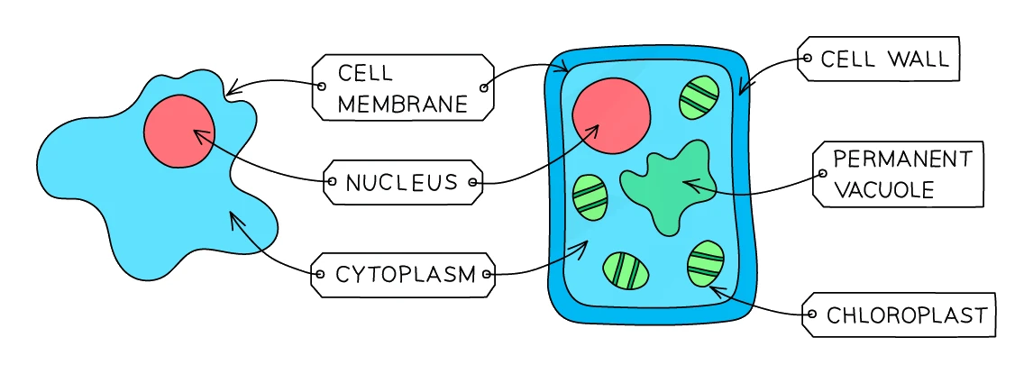
Plants need three extra structures that animals do not:
- A cell wall: plants do not have a skeleton, so each individual cell has to be rigid. The cellulose wall gives the plant body its shape and stops the cell from bursting when water rushes in.
- Chloroplasts: plants make their own food by photosynthesis, so they need an organelle that contains chlorophyll to capture light energy. Animals are heterotrophs and eat ready-made food, so they have no need for chloroplasts.
- A large permanent vacuole: plants control their support hydraulically. A full vacuole pushes outwards on the cell wall, making the cell turgid and keeping the plant standing up. When the vacuole loses water, the cell becomes flaccid and the plant wilts.
Common exam question
Which structures animal, plant and bacterial cells share
Question: From a labelled cell or a table of rows, choose which structures would also be found in an animal cell, which are found in all bacteria and all eukaryotic cells, or which row is correct for a red blood cell (1 mark).
Asked in 4 of the 23 papers, always as a multiple-choice part. The reasoning is the same each time: every cell has a cell membrane and cytoplasm; a cell wall and chloroplasts are never in an animal cell, so a cell wall is not something all eukaryotic cells share; a large vacuole is missing from animal cells too (it was the structure a white blood cell lacks). Watch the red blood cell row: it has cytoplasm but no nucleus and no cell wall.
Storage carbohydrate is tested the same way: a root cell stores starch, because plant cells do not make glycogen; animal cells store glycogen.
4. Prokaryotic cells
Bacterial cells (prokaryotes)
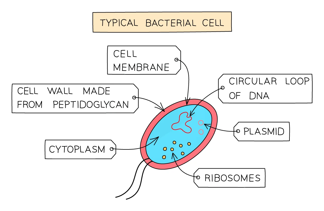
Bacteria are very different from animal and plant cells. They are prokaryotic, which means they have no and no membrane-bound at all. The main features of a bacterial cell are:
- A made of peptidoglycan (not cellulose; this is different from a plant cell wall)
- A just inside the wall
- filling the inside of the cell
- A single circular chromosome of DNA floating free in the cytoplasm
- Smaller circular pieces of DNA called plasmids, which can carry useful genes such as antibiotic resistance and which bacteria can swap between each other
- (smaller than the ribosomes in eukaryotic cells) for making proteins
- Some bacteria have a flagellum for swimming and pili for sticking to surfaces
Bacteria are also much smaller than eukaryotic cells. A typical bacterium is around 1 to 10 μm across, compared with 10 to 100 μm for an animal or plant cell.
Exam tip
Describing a bacterial cell in exam vocabulary
A bacterium has no nucleus, so never write "nucleus" for one: its DNA is a single circular chromosome lying in the cytoplasm ("nucleoid" is also accepted), and the small rings of DNA are plasmids. The two are credited as separate points, so name both. The wall is peptidoglycan, not cellulose, and there are no mitochondria or chloroplasts.
Yeast cells: a useful comparison
Yeast is a single-celled fungus (so it is a eukaryote, not a prokaryote). Yeast cells are roughly oval-shaped and contain:
- A with chromosomes
- (yeast respires both aerobically and anaerobically)
- A made of chitin (the same material as in multicellular fungi, different from both plant cellulose and bacterial peptidoglycan)
Yeast is important in two big industries: it makes carbon dioxide in bread baking (which makes the dough rise) and ethanol in brewing (anaerobic respiration of yeast on sugar produces alcohol).
5. Cell differentiation and specialised cells
What differentiation means
Differentiation is the process by which an unspecialised cell takes on the shape, mix and behaviour needed to perform one specific role in the body
When an embryo first forms, all of its cells look more or less the same. As the organism develops, each cell switches on a different set of genes and starts to change shape, change the organelles it contains, and produce different proteins. The result is a cell that is specialised for one specific job. A muscle cell is full of mitochondria; a nerve cell is long and thin; a sperm cell has a tail.
Two important points about differentiation:
- In animals, cells finish their differentiation very early on, mostly during embryonic and childhood growth. After that, only a small number of adult tucked away in particular tissues (bone marrow, skin, gut lining) still have the ability to differentiate; their job is to replace and repair damaged cells.
- In plants, many cell types keep the ability to differentiate throughout life. This is why a gardener can take a cutting from a shoot and grow a whole new plant: the cells in the cutting can still re-specialise into roots, leaves and so on.
Specialised cells you need to know
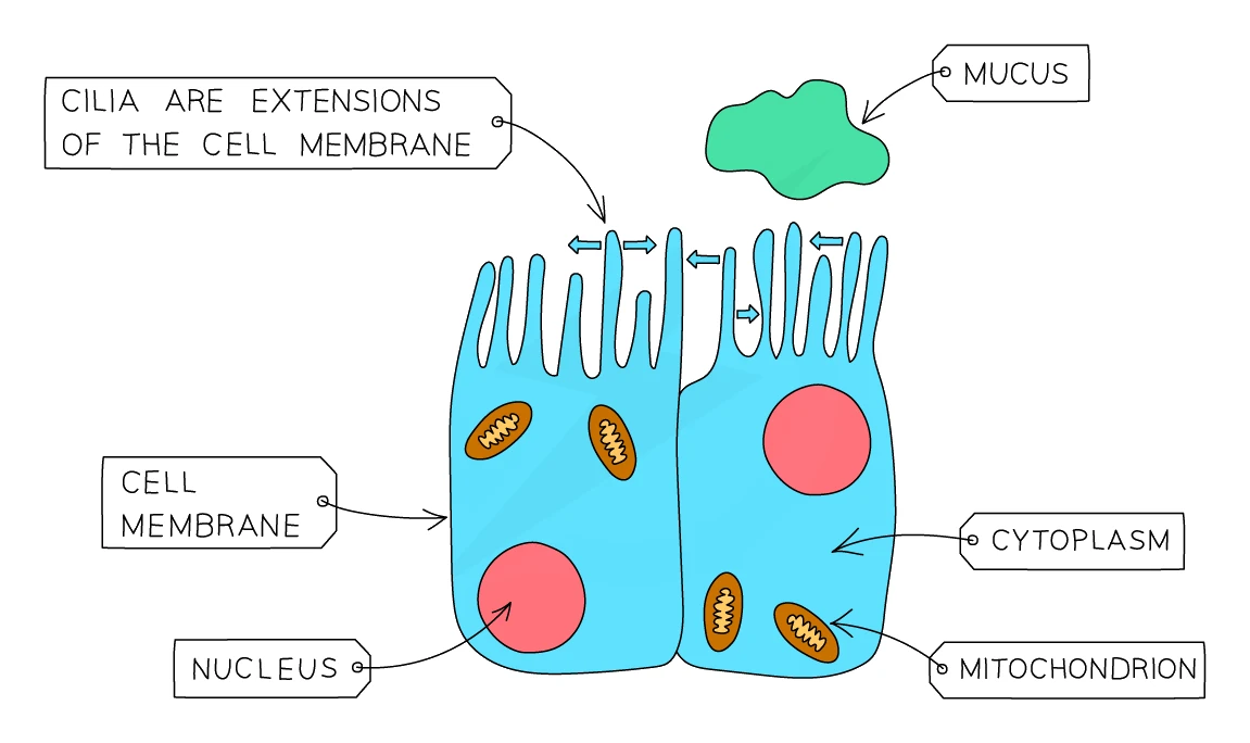
Ciliated epithelial (lining the trachea and bronchi)
- Has tiny hair-like extensions called cilia on its upper surface
- The cilia beat in a wave-like motion to sweep mucus (and the dust and microbes trapped in it) up the airways towards the throat, where it can be swallowed
- Helps keep the lungs clear of dirt and pathogens
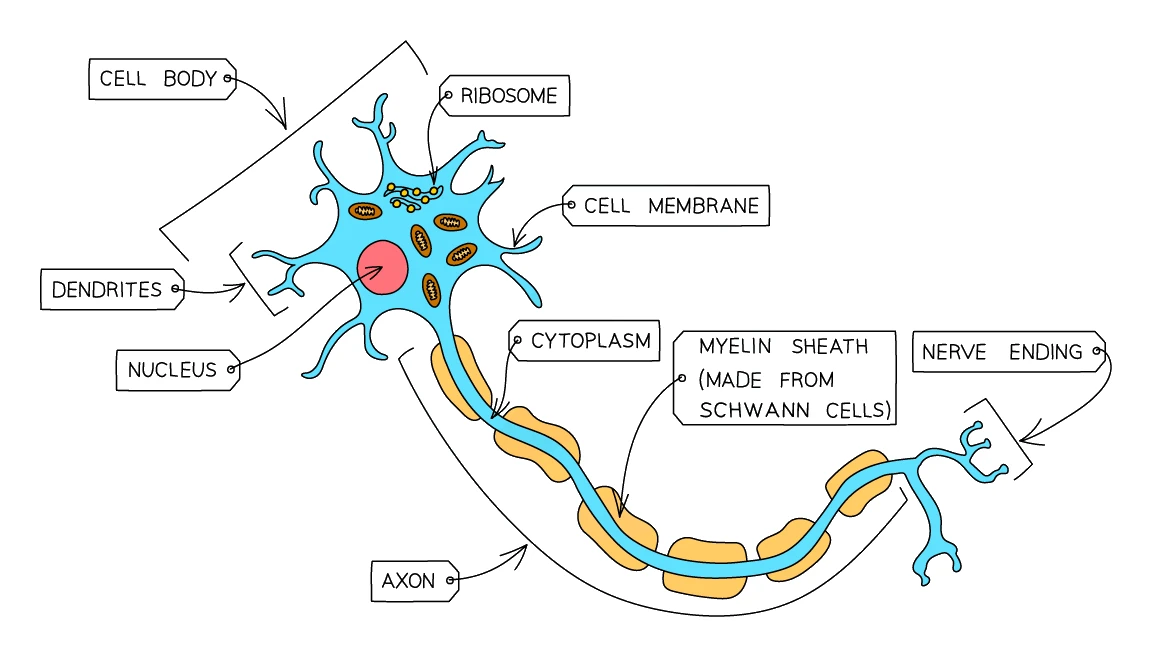
Nerve cell (neurone)
- Very long (some neurones in the leg are over a metre in length) so it can carry electrical signals over long distances
- Has many fine branches at each end called dendrites that connect to many other cells
- The long axon is wrapped in a fatty myelin sheath that insulates it and speeds up signal transmission
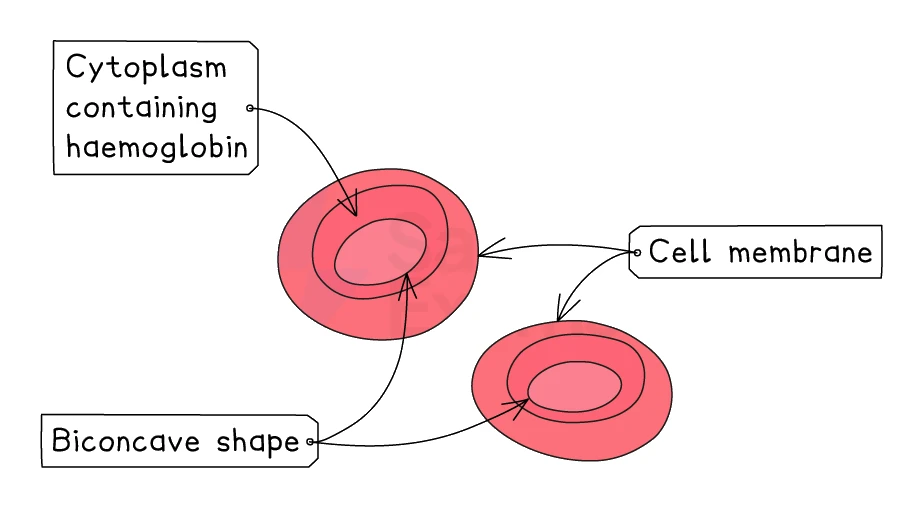
Red blood cell (erythrocyte)
- Biconcave disc shape (squashed in the middle on both sides), giving a high surface-area-to-volume ratio for efficient oxygen exchange
- No nucleus in mature red blood cells, leaving more room for haemoglobin
- Loaded with the oxygen-binding protein haemoglobin, which picks up oxygen in the lungs and lets go of it again wherever the body needs it
- Flexible enough to squeeze through capillaries narrower than itself
Sperm cell
- Has a long tail (flagellum) that beats to swim towards the egg
- Many mitochondria in the middle section to supply the energy needed for swimming
- The head contains a haploid nucleus with the male DNA, and the tip carries enzymes (in the acrosome) to digest the egg's outer layer
Egg cell (ovum)
- One of the largest cells in the human body
- Cytoplasm packed with nutrients to feed the developing embryo if fertilisation occurs
- After one sperm has entered, the membrane hardens to block any others
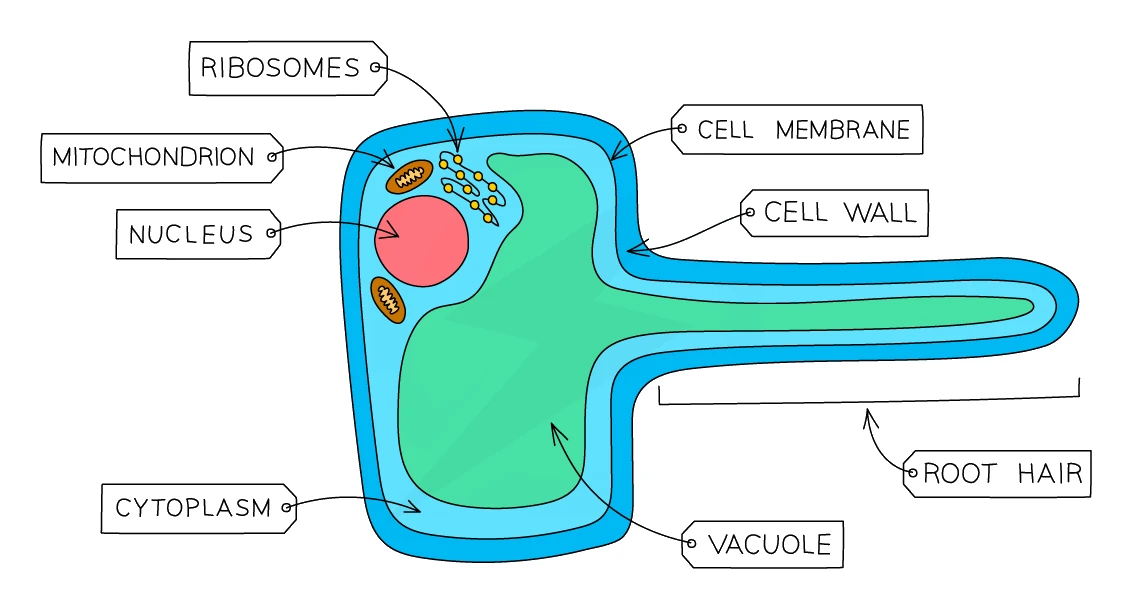
Root hair cell (in plant roots)
- Has a long extension sticking out into the soil that hugely increases its surface area for absorbing water and dissolved mineral ions
- A thin wall so that water moves quickly through it
- No (roots are underground, where there is no light)
- Found in their thousands in the root hair zone just behind the root tip
Common exam question
How a root hair cell is adapted for absorption
Question: Explain how a root hair cell is adapted for its function, or explain its role in absorbing water from the soil (3–4 marks).
Set in 2 of the 23 papers, both on Paper 2. Structure first, then process: the cell has a long extension that reaches into the soil, which increases the surface area; water is absorbed by osmosis, because the water potential inside the cell is lower than in the soil water; mineral ions are absorbed as well, by active transport. Each of those is a separate mark, so "large surface area for absorption" on its own earns one mark, not three. Linking the water to its use (photosynthesis, keeping cells turgid) or to the gradient set up by water loss from the leaves can also score.
Palisade mesophyll (in plant leaves)
- Found in the upper layer of the leaf, just under the epidermis, where the light is brightest
- Packed with many for photosynthesis
- Column-shaped and stacked tightly together side by side, so that as much chloroplast as possible can intercept the incoming light
6. Stem cells
What a stem cell is
A stem cell is an undifferentiated cell that can divide by mitosis to produce more stem cells, and that can also differentiate into specialised cells
Two key abilities make stem cells special:
- Self-renewal: they can keep dividing indefinitely to produce more stem cells
- Differentiation: when given the right chemical signals, they can develop into specialised cell types
Common exam question
Describing how stem cells differ from other cells
Question: Describe how stem cells differ from other body cells, or explain why stem cells can produce large numbers of one type of cell (2 marks).
Set in 3 of the 23 papers, all on Paper 2. The two marks come from three ideas in the definition: stem cells are undifferentiated (unspecialised), they can differentiate into other, specialised cell types, and they keep dividing by mitosis. Any two of the three score, so write all three. "They can become any cell" is accepted for the differentiation point. Producing large numbers of one cell type needs both halves: the stem cells divide repeatedly, and the new cells then specialise into the cell required.
Two types: embryonic and adult
| Embryonic stem cells | Adult stem cells | |
|---|---|---|
| Source | Very early embryos (a few days old) | Certain tissues in the adult body (bone marrow, gut lining, skin) |
| What they can become | Any cell type in the body. They are pluripotent | A limited range of cell types, usually closely related to the tissue they came from. Bone marrow stem cells, for example, can only become blood cells |
| Availability | Limited (need an embryo) | More accessible (can be taken from a living donor or the patient themselves) |
| Ethical issues | Major: many people view the destruction of an embryo as the destruction of a potential human life | Few ethical issues |
Uses of stem cells in medicine
Stem cells offer the possibility of replacing damaged cells in diseases where the body's own cells have stopped working properly. This is called therapy. Real and proposed applications include:
- Bone marrow transplants for treating leukaemia (a cancer of the blood). Healthy bone marrow stem cells from a donor are transplanted into the patient and produce new, healthy blood cells.
- Using stem cells to grow new skin for severe burns victims
- Treating Parkinson's disease by replacing damaged nerve cells in the brain
- Repairing spinal cord injuries by growing new nerve tissue
- Replacing damaged retinal cells in some forms of blindness
- Growing new heart muscle after a heart attack
Common exam question
Why bone marrow stem cells can treat blood disorders
Question: Explain how, or why, stem cells from bone marrow can be used to treat a range of different blood conditions (3 marks).
Set in 2 of the 23 papers, both on Paper 2. The general point comes first: blood stem cells can differentiate into every type of blood cell (red cells, white cells and platelets), worth one mark in the older scheme and two in the newer one (differentiating, then the range of cells). The other marks come from naming a cell type together with its job: red blood cells to carry oxygen, white blood cells to destroy pathogens or make antibodies, platelets to clot the blood. In the newer scheme "to treat anaemia" or "to treat leukaemia" in place of the job is ignored. If asked why a patient's own stem cells beat a donor's: same antigens (genetically identical), so no rejection and no immunosuppressant drugs, and no disease passed across.
Advantages of stem cell therapy
- Can treat conditions that have no other cure (paralysis, certain blindness, some cancers)
- Can replace cells exactly, restoring full function instead of just managing symptoms
- If the patient's own stem cells are used, there is no risk of rejection because the new cells carry the same antigens as the patient
Disadvantages and risks
- Ethical concerns about embryonic stem cells, since their use involves destroying a human embryo
- Risk of rejection if donor stem cells are used. The recipient is put on immunosuppressant drugs that stop their immune system from attacking the transplanted cells, but this leaves them more open to infections
- Risk of cancer: stem cells divide rapidly, and if they continue to divide uncontrollably after transplantation they can form a tumour
- Risk of viral infection carried over from the donor cells
- The technology is still expensive and experimental for most conditions
7. Microscopy and magnification
Why microscopes are needed
Most cells are far too small to see with the naked eye. A human red blood cell is only about 8 μm across; a bacterium is around 2 μm. A microscope uses lenses (or a beam of electrons) to magnify the image so structures invisibly small can be seen.
Light microscope vs electron microscope
| Light microscope | Electron microscope | |
|---|---|---|
| Source used to see the sample | Visible light through glass lenses | A beam of electrons through magnetic lenses |
| Maximum useful magnification | About ×1500 | Up to ×500 000 or more |
| Resolution (smallest detail visible) | About 200 nm | Down to 0.2 nm |
| Living specimens? | Yes, can view living cells in colour | No, the sample must be in a vacuum and is killed |
| Cost and size | Cheap and benchtop-sized | Very expensive, room-sized |
| What you can see | Cell wall, cell membrane, nucleus, chloroplasts, large vacuole | Also small organelles such as ribosomes and mitochondria in detail |
A light microscope is what you use in a school biology lab. An electron microscope is needed to see the smallest organelles (ribosomes, the internal structure of mitochondria, individual viruses).
The magnification equation
The relationship between the actual size of an object, the size of its image, and the magnification of the microscope is:
= image size ÷ actual size
Rearranged forms:
actual size = image size ÷ magnification
image size = actual size × magnification
Always make sure both sizes are in the same unit before dividing. Useful conversions:
1 mm = 1000 μm
1 μm = 1000 nm
1 mm = 1 000 000 nm
Common exam question
Calculating the magnification of a drawing or photograph
Question: The actual size of a cell, organism or virus is given in micrometres or millimetres; measure the printed image, often along a marked line, and calculate or determine its magnification (2–3 marks).
Set in 9 of the 23 papers. A 3-mark version usually follows the method: one for measuring the line with a ruler (a millimetre or two either side is allowed, and some schemes want the unit written with the reading), one for converting so both lengths share a unit (millimetres × 1000 gives micrometres), and the last for dividing image size by actual size. A 2-mark version gives one mark for the measurement or the division. A correct answer inside the accepted range earns full marks even with no working, and a wrong measurement carried correctly through the method still collects the method marks.
Magnification is a ratio: write it as "× 400", with no unit. Units written after it are ignored rather than penalised.
Worked example
Calculating magnification from a diagram
A diagram shows a palisade cell. You measure the line A–B on the diagram and find it is 30 mm long. The actual length of the cell from A to B is 150 μm. Calculate the magnification of the diagram.
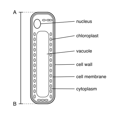
Solution:
- Convert the image measurement to the same unit as the actual size: 30 mm × 1000 = 30 000 μm
- Apply the formula: magnification = image size ÷ actual size
- magnification = 30 000 μm ÷ 150 μm = × 200