1. Activity and decay
What activity means
- A radioactive source is any object containing unstable nuclei that emit ionising radiation as they decay
- The of a source is defined as:
the rate at which the unstable nuclei in the source decay
- Activity is measured in becquerels (Bq), where:
1 Bq = 1 decay per second (one unstable nucleus in the source breaks down each second)
- A source with an activity of 1000 Bq has 1000 nuclei decaying per second; a source of 1 MBq has a million decays per second
Common exam question
Giving the unit of activity
Question: Give the unit of activity, or pick it out from a list of units (1 mark).
Asked in 4 of the 24 papers, twice as a multiple-choice item. The answer is the becquerel (Bq): one decay per second. Any prefix is accepted (kBq, for example), and so is a recognisable misspelling; one scheme also allowed the curie. The multiple-choice distractors are other SI units (the kilogram, newton, pascal, coulomb, joule and watt), so you only need to recognise the one unit that counts decays.
Activity decreases over time
- Every time a nucleus decays, it disappears from the pool of unstable nuclei
- So the number of unstable nuclei left in the sample is always falling
- Fewer unstable nuclei means fewer decays per second, so the falls with time as well
- The fall is not linear; it is faster at the start (when many unstable nuclei are present) and slower as time goes on. The shape is an curve
Decay is random
- Radioactive decay is a random process: it is impossible to predict which nucleus will decay next, or exactly when any one nucleus will break down
- All you can say is the probability that a given nucleus will decay in the next second
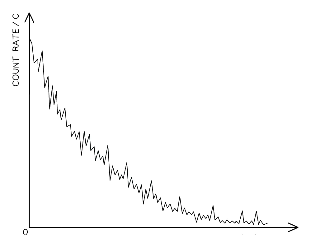
- The randomness can be seen by measuring the of a source using a Geiger–Müller (GM) tube
- The count rate is the number of decays the detector registers per second. When count rate is plotted against time the trace fluctuates up and down rather than following a perfectly smooth curve
- These fluctuations are direct evidence that decay is random. If decay were a steady, clockwork process the count rate would lie exactly on a smooth curve
Activity vs count rate (don't mix them up)
- Activity = rate at which unstable nuclei in the source actually decay (Bq)
- Count rate = rate at which the detector registers radiation
- Count rate is usually smaller than activity, because:
- The detector does not surround the source completely, so much of the radiation flies off and misses it
- Some radiation is absorbed by air, the GM tube wall, or any object in between
- Count rate is the practical, measurable quantity; activity is the theoretical quantity
2. Half-life
Definition
- Because individual decays are random, you cannot say "this particular nucleus will decay at 3:42 pm"
- What you can measure precisely is how long it takes for a large sample to halve. This is the half-life:
the of a radioactive isotope is the time taken for the number of unstable nuclei in a sample to fall to half of its original value
- Equivalently, since activity is proportional to the number of unstable nuclei, half-life is the time for the activity of the sample to drop to half its starting value
- Half-life is a property of the isotope itself. Every nucleus of a given isotope has the same probability of decaying per second, so the half-life is constant, and temperature, pressure and chemistry have no effect
Common exam question
Defining half-life
Question: State, describe or explain what is meant by the term half-life (2 marks).
Asked in 6 of the 24 papers, once as a multiple-choice item. The two marks are two separate ideas: the time taken ("how long it takes" is accepted; one scheme writes "average time") for the activity to halve, or for half of the unstable nuclei to decay. Count rate is accepted for activity and atoms for nuclei. Some schemes also allow the mass of the isotope, but one ignores it, so activity or nuclei is the safe choice.
Four of the five written schemes explicitly reject "half the time", so begin with "the time taken for". Name what halves: "the substance" or "the sample" is ignored, so say the activity, the count rate or the number of nuclei. A time with nothing halving, or a halving with no time, earns one mark of the two.
Half-lives vary enormously
- Different isotopes have wildly different half-lives:
| Isotope | Half-life | Use |
|---|---|---|
| Polonium-214 | ≈ 0.0002 s | Found in radon decay chains |
| Technetium-99m | ≈ 6 hours | Medical tracer |
| Iodine-131 | ≈ 8 days | Treating thyroid cancer |
| Carbon-14 | 5700 years | Carbon dating |
| Uranium-235 | 704 million years | Nuclear fuel |
| Uranium-238 | 4.5 billion years | Dating rocks |
- Short half-lives mean a very high for a short time; long half-lives mean a much lower activity but lasting far longer than any human timescale
Halving step by step
- After each half-life, the number of unstable nuclei (and the activity) is halved:
| Number of half-lives elapsed | Fraction of original isotope remaining |
|---|---|
| 0 | 1 (100%) |
| 1 | 1/2 (50%) |
| 2 | 1/4 (25%) |
| 3 | 1/8 (12.5%) |
| 4 | 1/16 (6.25%) |
| 5 | 1/32 (3.125%) |
- The pattern: after n half-lives, the fraction remaining is (1/2)ⁿ
Common exam question
Calculating an activity or a time with half-lives
Question: Given the half-life, calculate the activity after a stated time, show that a stated time is needed to fall below a given activity, or show the ratio of daughter to parent nuclei (3–5 marks; also set as multiple choice).
Set in 5 of the 24 papers. Write every halving down. In the three-mark version one mark is for halving the starting activity, one for showing the right number of half-lives (write the division, such as 36 ÷ 9 = 4) and one for the final value. For a "show that" time, halve until below the target, then turn that number of half-lives into years. For a ratio, find the parent nuclei left, subtract from the original total for the daughter nuclei (the total stays constant) and divide.
If a multiple-choice time is only half a half-life, more than half of the atoms remain: the answer lies between the starting number and half of it.
Worked example
Activity remaining after several half-lives
A radioactive source has an initial activity of 960 Bq. Its half-life is 4 hours. Find the activity after 20 hours.
Solution:
- Number of half-lives elapsed: 20 ÷ 4 = 5
- Halve the activity once for each half-life:
- After 1 half-life (4 h): 960 ÷ 2 = 480 Bq
- After 2 half-lives (8 h): 480 ÷ 2 = 240 Bq
- After 3 half-lives (12 h): 240 ÷ 2 = 120 Bq
- After 4 half-lives (16 h): 120 ÷ 2 = 60 Bq
- After 5 half-lives (20 h): 60 ÷ 2 = 30 Bq
Common exam question
Sketching the decay curve over three half-lives
Question: On the axes, sketch the decay of the sample during its first three half-lives, or draw crosses showing the number of atoms left after each of three half-lives (3 marks).
Set in 2 of the 24 papers. Most of the marks are for specific points, not a general shape. The sketch must start at the given initial activity at time zero, pass through half of it at one half-life (a plotted point is accepted as the evidence) and be a smooth curve that falls ever more slowly, so a straight line cannot earn the shape mark. For the crosses, each is a mark: at one, two and three half-lives, at a half, a quarter and an eighth of the starting number. If the next part asks for a curve of best fit through your crosses, the crosses may be read off that curve, so draw it smoothly through all of them.
Reading a half-life from a graph
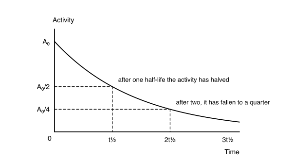
- To find the half-life from an activity-time graph:
- Read off the initial A₀ from the y-axis at t = 0
- Halve it to get A₀/2
- Draw a horizontal line from A₀/2 across to the curve, then drop straight down to the time axis
- The reading on the time axis is the
- It is a good idea to do this a second time (read at A₀/4) and divide the resulting time by 2 to check; you should get the same answer
Common exam question
Finding the half-life from a decay graph
Question: Use the graph to determine the half-life, or use a curve of best fit to estimate the time for the amount left to fall to a stated value (2 marks).
Asked in 6 of the 24 papers. One mark is for showing your method: a line across from half the starting activity (or count rate), or from the stated value, to the curve and down to the time axis, or the halving written in your working. Any halving along the curve is accepted, though the starting value is easiest. The second mark is for the reading, accepted within a small tolerance, so read carefully, with the axis unit.
When the points are given, draw the smooth curve of best fit first: the reading mark then follows your own curve even if it is slightly off. If asked why the points do not lie exactly on the curve, say that decay is random (a varying background count is also accepted).
Worked example
Reading a half-life from a decay curve
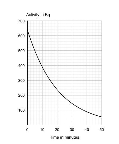
The graph is the decay curve of a radioactive sample whose activity at the start is 640 Bq. Use it to find the half-life of the sample.
Solution:
- Half of the starting activity: 640 ÷ 2 = 320 Bq
- Draw a line across from 320 Bq to the curve, then straight down to the time axis
- Read the time: the line meets the axis 2 small squares after the 10-minute line, and each small square is 2 minutes, so the time is 10 + 2 × 2 = 14 minutes
- Check with a quarter of the starting activity: 640 ÷ 4 = 160 Bq is reached at 28 minutes, and 28 ÷ 2 = 14 minutes
- Half-life = 14 minutes
3. Uses of radioactivity
The choice of radiation for any application depends on its range (how far it travels and what it can penetrate) and its ionising power.
Smoke detectors: alpha
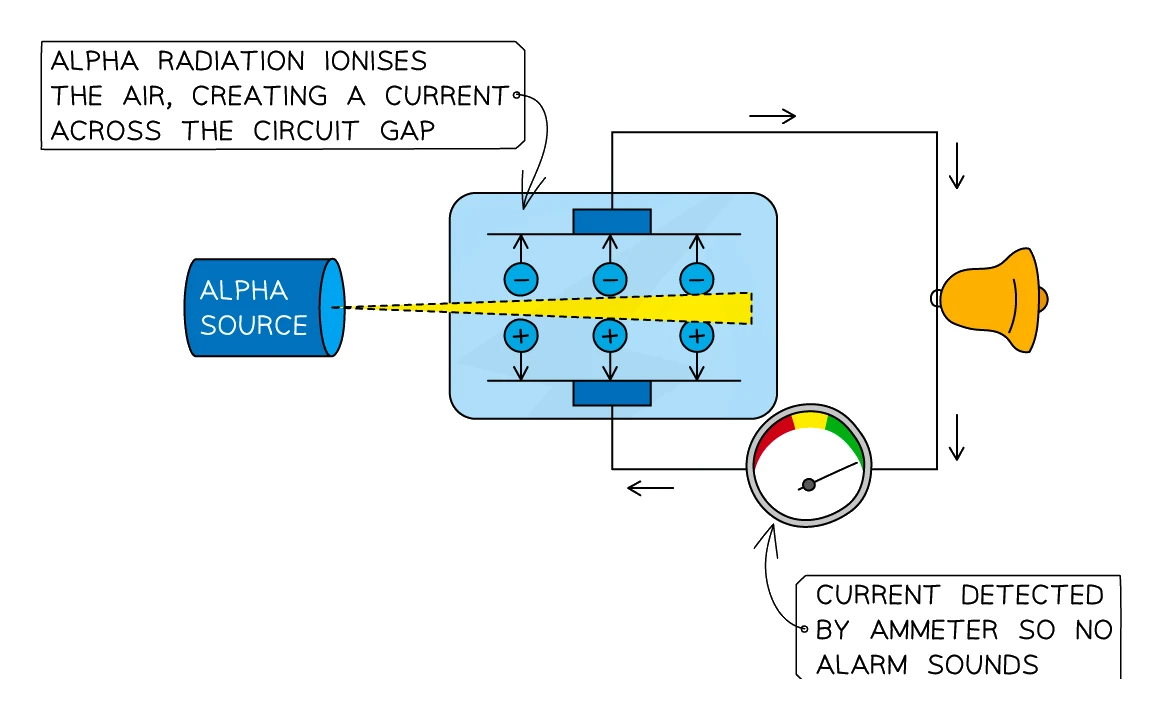
- A small alpha source (typically americium-241) sits inside the detector
- Inside a small chamber, the alpha particles ionise the air between two electrodes, creating a steady tiny current that flows through a circuit
- When smoke enters the chamber, the smoke particles absorb the alpha radiation before it can ionise the air
- The current drops sharply, and the microchip triggers the alarm
Why alpha is the right choice:
- Alpha is the most strongly ionising radiation, so it creates plenty of ions in the chamber and a strong baseline current
- Alpha is the least penetrating, so it cannot escape the detector, meaning there is no radiation hazard to the person standing under it
- Smoke is dense enough to absorb alpha but not beta or gamma. Beta and gamma radiation would slip through any normal puff of smoke unchanged, so the current would never drop and the alarm would never trip
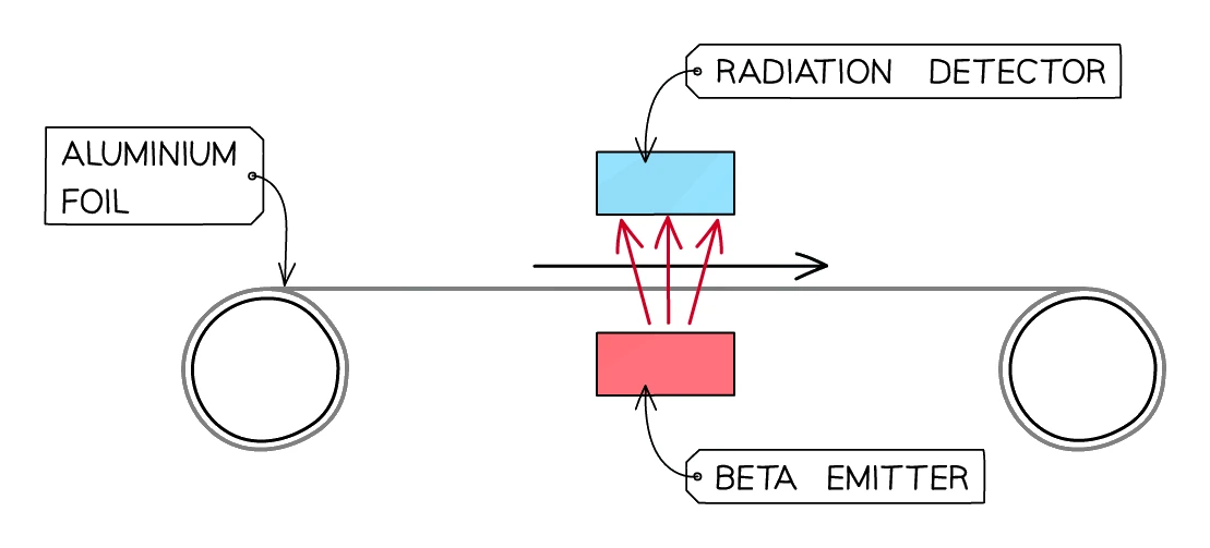
Measuring the thickness of materials: beta
- A beta source is placed on one side of a moving sheet of paper, cardboard or thin aluminium foil; a GM tube sits on the other side
- Some beta particles are absorbed by the sheet; the rest pass through and are counted by the GM tube
- If the sheet is too thick, more beta particles are absorbed and the count rate falls
- If the sheet is too thin, fewer beta particles are absorbed and the count rate rises
- A control system reads the count rate and adjusts the rollers to keep the thickness constant
Why beta is the right choice:
- Alpha would be stopped completely by even the thinnest paper, so the count rate would be zero no matter what thickness the sheet was
- Gamma would pass through almost unchanged, so changes in thickness would not register
- Beta is partially absorbed, which is exactly what is needed for the count rate to be sensitive to small changes in thickness
- The source must have a long (typically strontium-90, 28 years) so the count rate does not drift during the working life of the gauge
Treating cancer: gamma
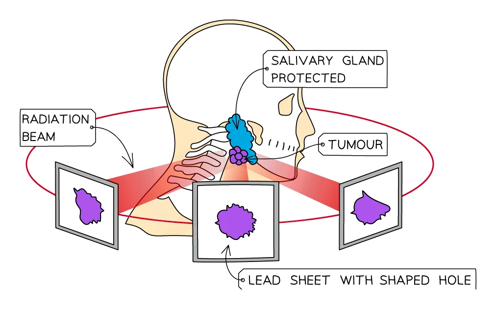
- Radiotherapy uses high-energy gamma rays directed at a cancerous tumour
- The gamma beam is aimed from several different directions, all crossing at the tumour. The healthy tissue along any one beam path receives only a small dose, but the tumour at the focus receives the full combined dose from every beam
- The beam is often produced by a rotating cobalt-60 source
Why gamma is the right choice:
- Gamma is highly penetrating, so it can reach a tumour deep inside the body from an external source
- Alpha and beta would be absorbed by the skin and surface tissue before reaching the tumour
Diagnosing cancer: tracers and PET scans
- A radioactive tracer is a small amount of a short-half-life radioisotope (often technetium-99m, half-life 6 hours) that is swallowed, inhaled or injected into the patient
- The tracer is chemically attached to a substance the body takes up, glucose for example. The substance carries the tracer to the organ being studied
- A detector (PET scanner) outside the body picks up the gamma emissions and builds up a map showing where the tracer has gone
- Bright spots show areas of high in the body. For example, fast-growing tumour cells take up far more glucose than healthy ones, so a tumour shows up as a bright spot
What makes a good tracer:
- Gamma emitter: gamma is the only radiation that can escape the body to reach the external detector
- Short half-life: the tracer should decay quickly so the patient is not radioactive for very long, but not so quickly that it decays before the scan is finished. A few hours is ideal
Sterilising medical equipment: gamma
- Surgical instruments are sealed inside their plastic packaging and exposed to a strong gamma source
- The gamma radiation kills any bacteria, viruses or spores on the instruments
- Because gamma is highly penetrating, it passes straight through the packaging. The instrument is sterilised without opening the seal, so it remains sterile until the surgeon opens the packet
- This method does not need high temperatures, so heat-sensitive instruments (plastics, electronics) can be sterilised this way
Irradiating food: gamma
- Food is exposed to gamma rays before being sold. The radiation kills bacteria, moulds and insects that would spoil the food or cause illness
- The food does not become radioactive (this is irradiation, not contamination; see Section 4)
- The food lasts longer on the shelf and is safer to eat
- Irradiated food is labelled with the Radura symbol, a stylised flower inside a broken circle
Carbon dating: beta
- Living things absorb carbon-14 continuously while they are alive, so the carbon-14 fraction in their tissues stays at the atmospheric level
- When the organism dies, no new carbon-14 is taken in. The carbon-14 already present decays (by beta emission) with a half-life of 5700 years
- The age of an ancient object (wood, bone, charcoal) can be worked out by measuring the remaining carbon-14 activity and counting how many half-lives have passed
- Useful for objects up to about 50 000 years old; for older rocks, uranium dating is used instead
4. Contamination and irradiation
These two words are commonly mixed up in exams. They describe completely different events.
Contamination
Contamination is the accidental transfer of a radioactive substance onto, or into, another object
- The contaminated object now physically contains radioactive atoms, usually in tiny amounts, on the surface or inside the body
- Because the radioactive atoms are part of the object, the object itself is now radioactive and will keep emitting radiation until those atoms decay away
- Typical examples:
- A radiation leak at a nuclear plant deposits radioactive dust on the ground and on workers' clothes
- Inhaling radioactive radon gas in a poorly ventilated cellar, where radon atoms enter the lungs and decay there
- Eating food grown on contaminated soil, so radioactive atoms enter the body
Irradiation
Irradiation is the process of exposing an object to ionising radiation from an external source
- The object is in the path of the radiation while the source is nearby, but no radioactive material is transferred onto or into it
- As soon as the source is removed or shielded, the irradiation stops, and the object is not radioactive afterwards
- Typical examples:
- A patient receiving a dental X-ray is irradiated while the X-ray machine is on, but does not become radioactive
- A worker in a hospital who walks past a shielded gamma source receives a small dose of irradiation
- Food being passed through a gamma sterilisation chamber
Side-by-side comparison
| Irradiation | Contamination | |
|---|---|---|
| What happens | The object is exposed to radiation from a separate source | A radioactive substance is transferred onto or into the object |
| Is the object radioactive after? | No, the radiation stops as soon as the source is removed | Yes, the object emits radiation until the radioactive atoms decay |
| Typical cause | Deliberate (X-ray, radiotherapy, sterilisation) | Accidental (leak, spill, ingestion, inhalation) |
| Type of radiation that matters most | Penetrating types (gamma, X-ray), because they reach you from outside | Ionising types (alpha), because once inside they damage cells most |
| How to prevent | Shielding (lead, concrete), distance, short exposure time | Sealing the source, gloves, airtight suits, no eating or drinking near sources |
| Easier to deal with? | Easier; remove the source and the problem stops | Harder; must remove or wait out every contaminated atom |
Half-life and which is more dangerous
- A short half-life means a high activity, with many decays per second per unit mass. Most of the radiation is emitted quickly
- This makes irradiation worse: while the source is active, anyone nearby is hit with a heavy stream of radiation
- For external doses (X-ray rooms, gamma sterilisers), short-half-life sources are the bigger danger
- A long half-life means a low activity but lasting for a very long time
- This makes contamination worse: a contaminated object stays radioactive for years or centuries and keeps irradiating its surroundings the whole time
- For environmental clean-up (nuclear leaks, buried waste), long-half-life isotopes are the bigger danger
5. Dangers of ionising radiation
How radiation harms cells
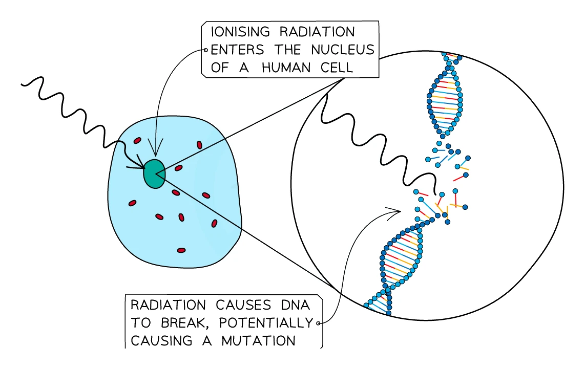
- All three types of nuclear radiation (and X-rays) are ionising: they knock electrons off atoms inside the body
- This can damage the DNA inside a cell in two main ways:
- Low doses: the DNA is damaged but not destroyed. The cell may repair the damage incorrectly, producing a mutation. A mutation in a gene that controls cell division can lead to cancer
- High doses: large numbers of cells are killed outright. Tissues stop working. Symptoms include radiation sickness (nausea, hair loss, burns) and, at very high doses, death
- Cells that divide quickly (bone marrow, gut lining, hair follicles, embryos) are most sensitive
- This is also why radiotherapy works: cancer cells divide faster than most healthy cells, so they are killed preferentially by a carefully aimed dose
Inside the body vs outside the body
- The dangers of alpha, beta and gamma flip around depending on whether the source is outside or inside the body:
| Outside the body | Inside the body | |
|---|---|---|
| Most dangerous | Gamma (and beta) | Alpha |
| Least dangerous | Alpha | Gamma |
| Why | Alpha is stopped by a few cm of air and by the skin. Beta and especially gamma can pass through clothing and skin and damage tissues underneath | Alpha is intensely ionising and deposits all of its energy in a tiny volume of tissue near where the atom decays, doing maximum damage. Gamma mostly passes straight through and exits the body |
- This is why alpha-emitting dust or gas is especially feared. Outside the body the alpha would barely reach the skin; inhaled or swallowed it irradiates the lungs or gut at point-blank range
- Polonium-210 (an alpha emitter) is a famous example: harmless if held in a sealed lump, lethal in microgram quantities if ingested
Common exam question
Explaining the hazard of an alpha emitter inside the body
Question: Explain the hazard of breathing in air containing radon, or discuss the hazards of a granite surface that releases radon, referring to contamination and irradiation (2–3 marks).
Set in 2 of the 24 papers. The credited points: radon is a gas, so it can be inhaled and get inside the body, which is contamination; its alpha particles then irradiate internal organs directly, with no dead layer of skin in the way; alpha is the most ionising radiation; and the damage to cells causes mutations or cancer. Outside the body alpha is stopped by skin or a few centimetres of air, so the solid thorium locked in granite can only irradiate you; it cannot get inside.
Use the two words precisely: a radioactive gas that enters the body contaminates it, while a source outside the body irradiates it. Each distinct idea is one mark.
Sources of background radiation
- Everyone is exposed to background radiation all the time. It comes from:
- Radon gas seeping up from rocks (the single biggest contribution in the UK, especially in granite areas)
- Cosmic rays from the Sun and outer space
- Rocks and soil containing tiny amounts of uranium, thorium and potassium
- Food and drink containing trace amounts of naturally radioactive isotopes (potassium-40, carbon-14)
- Medical sources such as X-rays, CT scans and radiotherapy
- Nuclear power and weapons testing (a small but measurable contribution)
- Background radiation levels are low and most people receive a very small dose each year. The risk of cancer at these levels is tiny, but it is not zero, which is why exposure should be kept as low as reasonably achievable
6. Safe handling of radioactive sources
There are three guiding principles for reducing the dose anyone receives from a source: time, distance and shielding.
Time
- The shorter the time you spend near a source, the smaller the total dose you receive
- Workers handling sources keep their tasks brief and the source out of its container only as long as needed
Distance
- Radiation spreads out from a source, so the further away you are, the lower the intensity at your position
- For a point source, the intensity follows an inverse-square law: doubling the distance from the source divides the dose rate by 4
- That is why sources are handled with tongs rather than fingers; the extra distance dramatically reduces the dose to the hand
Shielding
- A barrier between the source and the worker absorbs much of the radiation
- The right shielding depends on the radiation:
- Alpha: a sheet of paper or even a few cm of air is enough
- Beta: a few mm of aluminium
- Gamma: several cm of lead, or thick concrete; never fully stopped, only reduced
- X-ray rooms have lead-lined walls and lead aprons for the patient; nuclear waste is stored in thick concrete-and-steel casks
Practical safety rules in a school or hospital
- Keep sources in a lead-lined box when not in use
- Use tongs to lift and move sources; never touch with bare hands
- Point the source away from people
- Wear gloves, lab coats and (where appropriate) lead aprons or full lead-lined suits
- Where the source is a powder, dust or gas, wear an airtight suit to prevent contamination, keeping the radioactive material out of the eyes, nose and mouth
- Record how long the source is out, and how it has been used
- Wear a film badge (dosemeter), see below, so that any over-exposure is detected
Common exam question
Stating a precaution for handling a radioactive source
Question: State or give one precaution the teacher should take to work safely with a radioactive source (1 mark).
Asked in 2 of the 24 papers. Any one named action scores: handle the source with tongs (or gloves), keep it at arm's length or otherwise as far from you as possible, point it away from people, keep it in its lead-lined box when not in use, keep the time it is out of the box short, or wear a lead apron or goggles. One scheme ignores answers about being in another room, so name a precaution about handling, distance, time or shielding rather than moving the experiment elsewhere.
The film badge / dosemeter
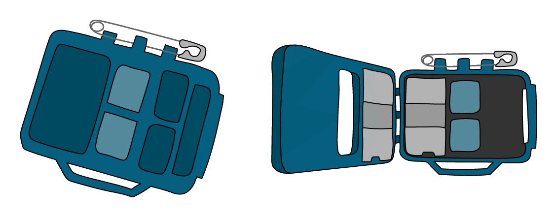
- A dosemeter is a small badge clipped to the clothing of anyone working with radiation
- The badge contains photographic film (or a modern equivalent), partly covered by strips of different absorber materials, typically paper, aluminium and lead, each of varying thickness
- Over a week or month the badge is removed and processed. The amount of darkening behind each absorber shows the dose of each type of radiation the wearer has received:
- Darkening behind no absorber but not behind the paper window ⇒ alpha (or low-energy radiation)
- Darkening behind paper but not aluminium ⇒ beta
- Darkening even behind lead ⇒ gamma or X-rays
- If the total dose exceeds the safe limit, the worker is taken off radiation work until the dose drops
7. Disposal of nuclear waste
Radioactive waste from power stations, hospitals and research labs has to be stored safely for as long as it remains active. How it is stored depends on the type of radiation and the .
Low-level waste
- Materials with only alpha or weak beta activity (used gloves, paper, syringes from hospitals)
- Sealed inside plastic bags or metal canisters and buried in shallow, monitored landfill sites
- The packaging easily absorbs the alpha and beta emissions
Intermediate-level waste
- More strongly active beta and gamma waste (reactor parts, contaminated tools)
- Sealed inside steel drums, often surrounded by concrete to form a heavy block
- Stored in concrete-lined silos above ground or in shallow burial sites
High-level waste
- Spent nuclear fuel rods and the products of fission
- Strongly gamma-emitting, with very long half-lives (thousands to millions of years)
- Storage strategy:
- First cooled in water-filled ponds for several years next to the reactor; water shields the gamma and removes the heat of decay
- Then encased in vitrified glass blocks (mixing the waste into molten glass that solidifies into a solid, leach-resistant form)
- Then sealed inside lead-lined, thick concrete casks
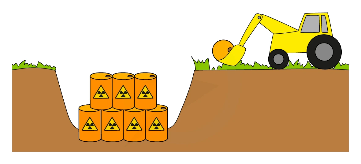
- Finally buried deep underground in geologically stable sites such as old salt mines or stable rock formations where there is no groundwater movement
Why it is so hard
- Storage must last as long as the waste is active. For some isotopes this is tens of thousands of years, far longer than any civilisation has lasted
- Containers must:
- Resist corrosion for centuries (stainless steel with copper outer layers; expensive to manufacture)
- Survive earthquakes, floods and accidental impact
- Stop water reaching the waste (water carries dissolved radioactive atoms into the environment)
- The chosen burial site must have:
- Low risk of earthquakes and other natural disasters
- No groundwater flow that could transport radioactivity to lakes, rivers or aquifers
- High security so the waste cannot be stolen or accidentally exposed
- For very low-activity liquid waste, dilution in large volumes of seawater is sometimes used. This works only because the radioactivity is spread so thinly that any single point in the sea is at or near background level; it relies on the radioactivity being too diffuse to harm anything, not on it disappearing