1. Why humans need a circulatory system
A human body contains around 37 trillion cells, most of them buried deep inside organs many centimetres from the body's surface. Every one of these cells needs a constant supply of oxygen, glucose, water, mineral ions, hormones and other essentials, and a fast way to get rid of waste carbon dioxide, urea and heat. Diffusion alone is far too slow to do this on a body-wide scale, so humans (and most other large animals) have a circulatory system: a closed loop of vessels through which blood is pumped by the heart.
The system has three parts:
- A pump (the heart)
- A network of vessels (arteries, capillaries, veins)
- A transport fluid (the blood)
Together they deliver substances to every cell in the body and carry the waste products away to the organs that excrete them.
2. The composition of blood
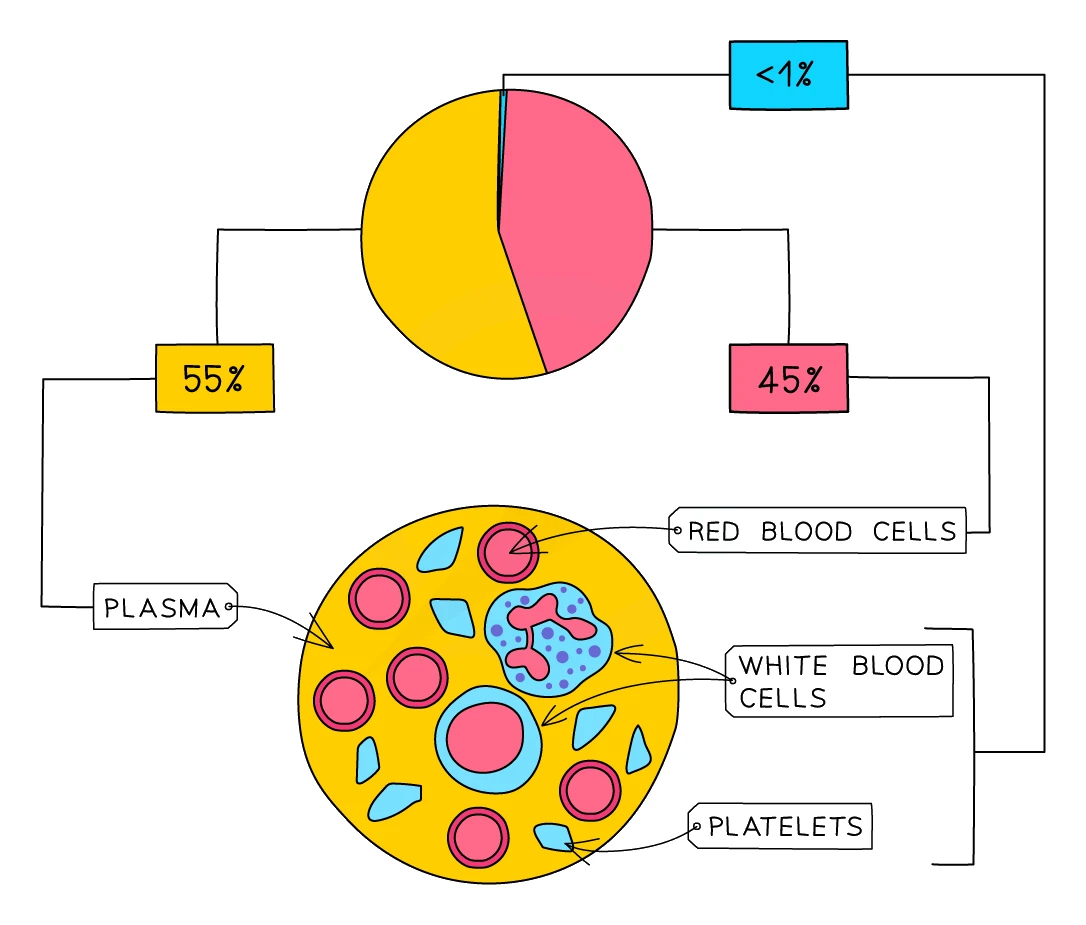
An adult human carries around 5 litres of blood. It is made up of four main components:
| Component | What it looks like | Roughly what fraction of blood |
|---|---|---|
| Plasma | Pale yellow watery liquid | About 55% by volume |
| Red blood cells | Tiny biconcave discs, no nucleus, red colour | About 45% by volume |
| White blood cells | Larger cells with a nucleus, less than 1% by volume | A small fraction |
| Platelets | Tiny fragments of cells, no nucleus | A small fraction |
Plasma
Plasma is the watery fluid the other components are suspended in. About 90% of it is water, with all sorts of useful substances dissolved in it:
- Carbon dioxide dissolved from respiring cells, being carried to the lungs to be breathed out
- Digested food molecules (glucose, amino acids, mineral ions) absorbed from the small intestine and being delivered to every cell
- Urea, a nitrogen-containing waste from the liver, being carried to the kidneys for excretion
- Hormones (chemical messengers) released by endocrine glands, being delivered to their target organs
- Heat energy released by respiring cells, being moved to cooler parts of the body or to the skin
Red blood cells (erythrocytes)
Red blood cells are highly specialised for one job: carrying oxygen from the lungs to every respiring cell in the body. Three adaptations make them excellent at this:
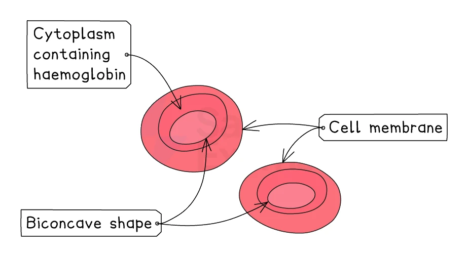
- Packed with , an iron-containing protein that binds reversibly with oxygen. In the oxygen-rich environment of the lungs, haemoglobin combines with oxygen to form . In the low-oxygen environment of a respiring tissue, oxyhaemoglobin releases its oxygen back to haemoglobin and the oxygen diffuses into the tissue.
- No nucleus, which leaves more room inside the cell for haemoglobin. One mature red blood cell holds about 250 million haemoglobin molecules.
- Biconcave disc shape (pinched in the middle on both sides). This gives a large surface-area-to-volume ratio for oxygen to diffuse in and out quickly, and lets the cells be flexible enough to squeeze through narrow capillaries.
A red blood cell only lives for about 4 months. Old ones are broken down in the liver, and new ones are made constantly in the bone marrow.
Common exam question
Comparing a red blood cell with another cell
Question: Give structural differences between a human red blood cell and a white blood cell or fish red cell, or explain why a spherical or fish red cell carries oxygen less efficiently (1–3 marks).
Set in 3 of the 23 papers. For structure, start with the nucleus: a human red cell has no nucleus, the one-mark difference from a fish red cell. Against a white blood cell, any three of these earn the 3 marks: no nucleus, a biconcave disc rather than an irregular shape, smaller, and contains haemoglobin. Name features, not jobs. For efficiency, the biconcave disc gives a larger surface area to volume ratio, so oxygen diffuses in and out faster; against the fish cell, having no nucleus leaves more room for haemoglobin; and the flexible shape squeezes through capillaries. Write each point as a comparison ("a larger ratio than the sphere has"), from either cell's side. One scheme ignores "gas exchange" on its own: name oxygen.
Common exam question
Explaining the effects of having fewer red blood cells
Question: Explain why someone with fewer red blood cells or less haemoglobin (anaemia, thalassaemia) feels tired, or comment on how the condition affects them (2–4 marks).
Asked in 2 of the 23 papers. The marks are a chain, and each link is a separate point: less haemoglobin, so less oxygen is transported (to the muscles), so less aerobic respiration and less energy or ATP released, so the person is tired or out of breath, or respires anaerobically and builds up lactic acid. Write every link in order: "less oxygen so they are tired" skips the respiration step, which carries a mark of its own. The same chain explains why a heart with narrowed coronary arteries copes less well with exercise and, run the other way, why a trained athlete can run faster.
White blood cells (leucocytes)
belong to the immune system. Their job is to defend the body against pathogens. Two main types exist:
- Phagocytes carry out non-specific immunity. They engulf and digest any pathogen they meet.
- carry out specific immunity. They produce antibodies, proteins that recognise one particular pathogen.
A typical white blood cell is much larger than a red blood cell and has a prominent nucleus when viewed under a microscope. Both types are covered in detail in section 6.
Platelets
Platelets are not whole cells but tiny fragments broken off from much larger cells in the bone marrow. They have no nucleus. Their job is to make the blood clot at a wound, which seals the cut, prevents excessive blood loss, and stops bacteria getting in. Covered in section 8.
3. Blood vessels
Humans use three kinds of blood vessel, each shaped for one job:
| Feature | Artery | Vein | Capillary |
|---|---|---|---|
| Direction of flow | Away from the heart | Towards the heart | Between artery and vein, through tissues |
| Blood pressure | High (the heart's pump has just pushed blood out) | Low (most of the pressure has been lost in the capillaries) | Low |
| Wall thickness | Thick, muscular, with elastic fibres | Thin | One cell thick |
| Lumen (the inner channel) | Narrow | Wide | Very narrow (only just wide enough for a single red blood cell) |
| Valves? | No (the semilunar valves at the start of the aorta and pulmonary artery belong to the heart, not to the arteries themselves) | Yes, frequent pocket-shaped valves | No |
| Blood content | Usually oxygenated (exception: pulmonary artery) | Usually deoxygenated (exception: pulmonary vein) | Both oxygenated and deoxygenated, depending on where in the body |
A handy memory trick: Arteries carry blood Away from the heart.
Why each vessel has the structure it does
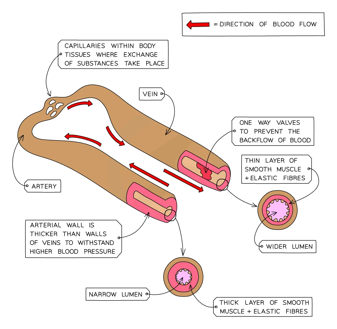
Arteries have to cope with the high pressure of blood freshly pumped from the heart:
- Thick muscular wall withstands the pressure without bursting
- Elastic fibres in the wall stretch when the heart beats and recoil between beats, evening out the surges of pressure
- Narrow lumen keeps the pressure high so blood is delivered quickly to distant tissues
Veins carry blood back to the heart under much lower pressure:
- Thinner wall is enough for the low pressure
- Wider lumen offers less resistance to the slow blood flow
- Valves stop the blood from flowing backwards. When you tense the muscles around a vein (e.g. by walking), the squeezing pushes blood forwards; the valves stop it sliding back when the muscle relaxes
- Many large veins run between skeletal muscles, so movement of the limbs helps pump the blood back to the heart
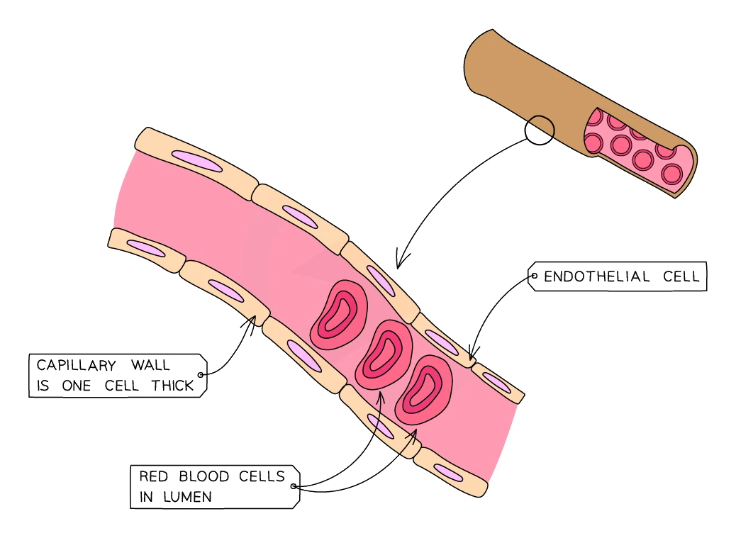
Capillaries are where the actual exchange between blood and tissues happens. They form a dense network branching out from arteries and joining back up into veins inside every tissue:
- One cell thick: the wall is a single layer of cells, so the diffusion distance for oxygen, glucose, CO₂ and urea is as short as possible
- Very narrow lumen forces red blood cells to pass through one at a time, slowing the flow and giving plenty of time for diffusion
- "Leaky" walls allow plasma to seep out into the surrounding tissue as tissue fluid, which bathes the cells and delivers their supplies. Most tissue fluid is reabsorbed; the rest is collected by a separate network of lymphatic vessels and returned to the bloodstream
Common exam question
Explaining how an artery wall differs from a vein wall
Question: Give, or explain, two differences between the structure of an artery (or the aorta) and a vein (or the vena cava) (2–4 marks).
Set in 2 of the 23 papers. Across the two schemes the credited differences are: the artery has a thicker wall, more muscle, more elastic fibres, a narrower lumen and no valves; the vein has the converse of each. In the 4-mark "explain" version the scheme marks in pairs, a structure point and its reason: a thick elastic layer because the pressure is higher and the wall must stretch and recoil to smooth the flow; a thick muscle layer to control blood flow; no valves because the pressure is enough to prevent backflow. "Thick wall because of high pressure" on its own is held to 2 marks, so name the elastic and muscle layers separately, each with its own reason.
4. The heart
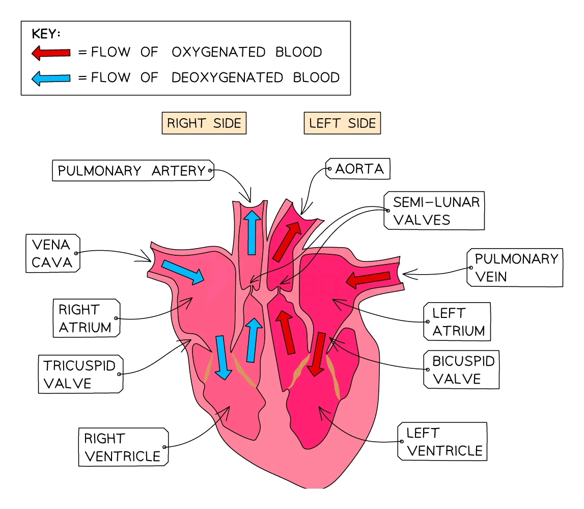
The heart is a muscular pump about the size of a closed fist, sitting just left of centre in the chest. It is really two pumps stuck together, working side by side: a right side that pumps deoxygenated blood to the lungs, and a left side that pumps oxygenated blood to the rest of the body.
Four chambers
Each side of the heart has two chambers:
| Chamber | What it does |
|---|---|
| Right atrium | Receives deoxygenated blood from the body via the vena cava |
| Right ventricle | Pumps that deoxygenated blood to the lungs via the pulmonary artery |
| Left atrium | Receives oxygenated blood from the lungs via the pulmonary vein |
| Left ventricle | Pumps that oxygenated blood to the whole body via the aorta |
The atria are the small upper chambers that receive blood from the veins. The ventricles are the larger lower chambers that pump blood out into the arteries.
The two sides are completely separated by a thick muscle wall called the septum. This separation keeps oxygenated and deoxygenated blood from mixing.
Common exam question
Answering questions on a labelled heart diagram
Question: On a labelled heart diagram, identify a chamber, vessel or the septum, or give two differences between the blood in two labelled vessels (1–2 marks each, often multiple choice).
Asked in 3 of the 23 papers, each time opening a longer heart question. The heart is drawn as if you were facing the person, so its left side is on the right of the page. Atria are above, ventricles below; the vena cava brings deoxygenated blood into the right atrium; the aorta leaves the left ventricle, which pumps oxygenated blood to the body; the septum is the thick wall between the ventricles.
For two labelled vessels, the marks are the two gases: the one carrying blood from the lungs or the heart's left side has more oxygen and less carbon dioxide. Give one gas per line, saying which vessel has more; pressure is not composition and earns nothing. The pulmonary artery carries deoxygenated blood, the pulmonary vein oxygenated.
Why the left ventricle has the thickest wall
The left ventricle is built with a much chunkier muscle wall than its right counterpart. The reason is workload: the left ventricle has to drive blood at high pressure all the way around the whole body (the systemic circuit), while the right ventricle only sends blood a short way to the lungs (the pulmonary circuit). A thicker, stronger muscle generates the higher pressure.
The right ventricle deliberately pumps at lower pressure: the delicate capillaries in the alveoli of the lungs would be damaged by high-pressure blood.
Common exam question
Explaining why the left ventricle wall is thicker
Question: Explain the differences between the wall of the left ventricle and the wall of the right ventricle (3 marks).
Asked in 2 of the 23 papers, both times for 3 marks from four available points: the left wall is much thicker, it has more muscle, so it generates more pressure (or force), because it pumps blood all around the body while the right ventricle pumps only to the lungs. "Thicker muscle" covers the first two points in one phrase, so add the pressure or the destination for the third mark. Say the wall generates or produces the higher pressure: "withstands high pressure" is ignored in both schemes. Writing the converse about the right ventricle (thinner, less muscle, less pressure, only to the lungs) is credited equally.
Four valves
The heart has four one-way valves that stop blood from flowing backwards:
| Valve | Where it is | What it does |
|---|---|---|
| Right atrioventricular valve (tricuspid) | Between right atrium and right ventricle | Stops blood flowing back from the right ventricle into the right atrium when the ventricle contracts |
| Left atrioventricular valve (bicuspid / mitral) | Between left atrium and left ventricle | Stops blood flowing back from the left ventricle into the left atrium |
| Semilunar valve (pulmonary) | At the start of the pulmonary artery | Stops blood flowing back from the pulmonary artery into the right ventricle |
| Semilunar valve (aortic) | At the start of the aorta | Stops blood flowing back from the aorta into the left ventricle |
The pathway of blood through the heart
Starting from where blood arrives back from the body:
- Deoxygenated blood from the body enters the right atrium through the vena cava.
- The right atrium contracts, pushing the blood through the right atrioventricular (tricuspid) valve into the right ventricle.
- The right ventricle contracts, pushing the blood through the pulmonary semilunar valve into the pulmonary artery.
- The pulmonary artery carries the deoxygenated blood to the lungs, where it picks up oxygen and releases carbon dioxide in the capillaries surrounding the alveoli.
- Oxygenated blood returns from the lungs through the pulmonary vein into the left atrium.
- The left atrium contracts, pushing the blood through the left atrioventricular (bicuspid / mitral) valve into the left ventricle.
- The left ventricle contracts strongly, pushing the blood through the aortic semilunar valve into the .
- The aorta carries the oxygenated blood out to the whole body.
In reality both atria contract together, then both ventricles contract together; the diagram describes one side at a time only for clarity.
The coronary arteries
The heart muscle itself needs a constant supply of oxygen and glucose for its own respiration. These are delivered by the coronary arteries, which branch off the aorta as soon as it leaves the heart and run across the outside of the heart muscle. If a coronary artery becomes blocked, the patch of heart muscle it feeds runs out of oxygen and dies, causing a heart attack (section 7).
5. The double circulation
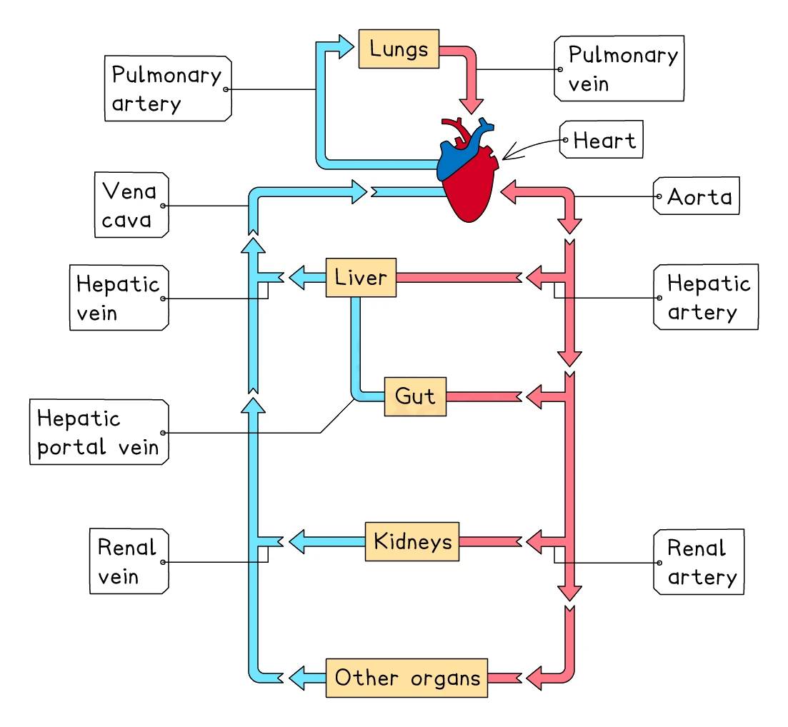
The mammalian circulation is a double circulation: blood passes through the heart twice for every complete journey around the body. The two halves of the loop are called the pulmonary circuit and the systemic circuit:
- Pulmonary circuit (heart → lungs → heart): the right side of the heart pumps deoxygenated blood out to the lungs through the pulmonary artery. Blood picks up oxygen and drops off CO₂ in the lungs, then returns to the left side of the heart through the pulmonary vein.
- Systemic circuit (heart → body → heart): the left side of the heart pumps oxygenated blood out to every other organ through the aorta. Cells take the oxygen and add their waste CO₂; the now-deoxygenated blood returns to the right side of the heart through the vena cava.
Why a double circulation is so useful
In a single circulation (like in a fish), blood passes through the heart only once and slows right down in the gills' capillaries before being pumped on to the rest of the body. By the time it reaches the muscles it is moving sluggishly. In a double circulation, the blood is re-pressurised between the lungs and the rest of the body, so it can be delivered fast to every tissue. This allows mammals to be much more active than fish of comparable size.
The main blood vessels of the major organs
| Organ | Vessel taking blood TO the organ | Vessel taking blood AWAY |
|---|---|---|
| Heart | Vena cava, pulmonary vein (these bring blood to the heart's chambers, not its muscle) | Aorta, pulmonary artery |
| Heart muscle itself | Coronary artery | Cardiac vein |
| Lungs | Pulmonary artery (deoxygenated) | Pulmonary vein (oxygenated) |
| Liver | Hepatic artery (oxygenated) and hepatic portal vein (from gut) | Hepatic vein |
| Kidneys | Renal artery | Renal vein |
6. White blood cells and the immune response
Two types, two strategies
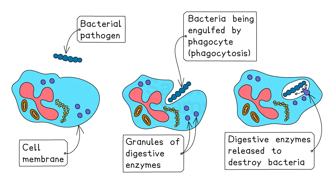
Phagocytes are the rapid-response part of the immune system. They patrol the blood and tissues looking for anything that does not belong (bacteria, viruses, fungi). When they find a pathogen they engulf it whole (a process called ) and digest it inside themselves using digestive enzymes. This is a non-specific response: a phagocyte will engulf any pathogen it meets, not just one type.
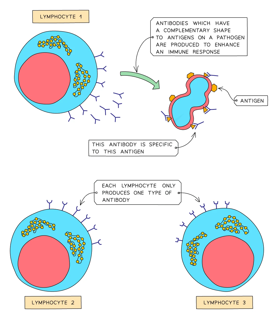
Lymphocytes are the targeted-response part of the immune system. They produce antibodies: proteins that exactly fit a specific feature on the surface of one type of pathogen. This is a specific response, since each lymphocyte makes antibodies against only one type of pathogen.
Antigens and antibodies
- An antigen is a molecule (usually a protein) found on the surface of a cell or pathogen. The body recognises antigens that belong to itself as "self", and any antigen that doesn't match as "foreign".
- An antibody is a Y-shaped protein, made by a lymphocyte, that has a complementary shape to one specific antigen. The antibody locks on to its matching antigen like a key fits its lock.
When antibodies bind to pathogens, they:
- Clump pathogens together (agglutination), making them easier for phagocytes to engulf in batches
- Mark pathogens for destruction, by giving the phagocytes a clear signal of what to engulf
- Neutralise toxins released by some bacteria, by binding to the toxin so it can no longer harm body cells
Common exam question
Describing how white blood cells fight a pathogen
Question: Describe how the immune system responds to disease, explain what an immune response is, or explain how antibodies protect against a pathogen (1–3 marks).
Asked in 4 of the 23 papers. The credited points: white blood cells respond to the antigens on a pathogen; phagocytes engulf and digest it; lymphocytes produce antibodies that are specific to that antigen and clump, label or burst the pathogen so it is destroyed; memory cells are made. Keep each cell with its own job. A swapped pairing ("lymphocytes engulf", "phagocytes produce antibodies") is rejected in one scheme; in another it keeps only the mark for naming a white blood cell and loses the mark for what it does. And when a one-mark part asks which cell makes antibodies, "white blood cells" is ignored and "phagocytes" is rejected. Only lymphocytes scores.
How the immune system fights an infection
- A pathogen enters the body and starts to multiply, releasing toxins and damaging cells.
- Phagocytes are the first to respond. They engulf and digest pathogens they encounter (non-specific phase).
- Eventually a meets a pathogen whose antigens match its receptors.
- That lymphocyte begins making specific antibodies against that antigen and divides rapidly, producing many identical lymphocytes that all churn out the same antibody.
- The antibodies bind to the antigens on the pathogens, clumping them and tagging them for destruction.
- Phagocytes engulf the tagged pathogens in bulk and digest them.
- Some of the lymphocytes remain in the body afterwards as memory cells, ready to mount a faster response if the same pathogen ever attacks again.
Common exam question
Describing how a phagocyte destroys a pathogen
Question: Describe the role of phagocytes, or describe what is happening at each stage as a phagocyte destroys a bacterium (3–4 marks).
Set in 2 of the 23 papers. The stages, each worth a mark: the phagocyte engulfs the pathogen (surrounds it with pseudopodia; "ingests" is allowed); the pathogen sits inside a vesicle, the phagosome; vesicles containing enzymes fuse with it; the enzymes digest the pathogen; the products are expelled from the cell. In the 3-mark version "digests it using enzymes" takes two of the three marks, so say enzymes, not just "digests". In the 4-mark version the vesicle and the fusion step carry marks of their own, so "engulfs and digests" cannot reach full marks.
Immunity and memory cells
The first time you meet a new pathogen, the immune response is slow (it takes the lymphocytes time to find the right one and multiply), and you feel ill. By the time the pathogen is cleared, the body has a stock of memory cells specific to that pathogen.
If the same pathogen invades a second time, the memory cells recognise it immediately and produce more antibodies, faster, and in greater quantities than the first time around. The pathogen is destroyed before it has a chance to cause symptoms. You are now immune to that pathogen.
Vaccination
Vaccination is a way of getting the memory-cell benefit without ever needing to suffer the disease. A vaccine contains a harmless version of a pathogen, prepared in one of three ways:
- Killed pathogens (e.g. the older type of flu vaccine)
- Weakened (attenuated) live pathogens (e.g. the MMR vaccine for measles, mumps, rubella)
- Pieces of pathogen such as surface antigens (e.g. the hepatitis B vaccine)
The vaccine carries the pathogen's without being able to cause the disease. The body responds as if it has been infected:
- Lymphocytes recognise the antigens.
- They produce specific antibodies and divide.
- Memory cells are formed and remain in the bloodstream.
If the real pathogen ever attacks later, the memory cells trigger a fast, strong antibody response and the person remains symptom-free. Vaccination has dramatically reduced or eradicated diseases including smallpox (eradicated worldwide), polio (almost eradicated), measles, rubella, tetanus and many others.
Common exam question
Explaining how vaccination protects against a disease
Question: Explain how a vaccine stops a person or animal developing a disease, or why vaccination is not used on someone whose immune system is suppressed (2–4 marks).
Set in 3 of the 23 papers. Build the answer in order: the vaccine contains a weakened or inactive form of the pathogen, which carries its antigens; lymphocytes recognise them and respond; memory cells are produced; so if the real pathogen arrives there is a secondary response, with antibodies made sooner, faster and in greater quantity. Each step is a separate mark. One scheme ignores any mention of phagocytes, so do not describe engulfing. For the suppressed-immune-system version reverse the chain: the person cannot produce antibodies or memory cells, so there is no secondary response, and a live vaccine could even give them the disease.
7. Coronary heart disease
Coronary heart disease (CHD) is the leading cause of death in many developed countries. It develops when the coronary arteries (the small arteries that supply the heart muscle itself) become narrowed by fatty deposits.
How it develops
- Cholesterol and other lipids in the blood get deposited on the inside walls of the coronary arteries, forming patches called atheromas (sometimes called fatty plaques).
- The atheromas gradually narrow the lumen of the artery, restricting the flow of blood to the heart muscle.
- The rough surface of an atheroma can trigger a blood clot to form. The clot blocks the artery further, or even completely.
Two outcomes
- Partial blockage: the heart muscle still gets some blood but not enough during exercise. This causes a sharp, gripping chest pain called angina, especially during physical exertion.
- Complete blockage: the patch of heart muscle fed by that artery is suddenly cut off from oxygen. Cells in that region cannot respire aerobically and quickly die. This is a heart attack (medically, a myocardial infarction). If a large enough area dies, or if the dying area includes the heart's electrical pacemaker, the heart may stop beating altogether.
Risk factors
Coronary heart disease has several risk factors that increase a person's chance of developing it. Most can be managed, given the right lifestyle choices:
| Risk factor | Why it raises the risk |
|---|---|
| High-fat diet (especially saturated fat and cholesterol) | Raises blood cholesterol, accelerating atheroma formation |
| High blood pressure (hypertension) | Increased force damages the artery walls, making it easier for atheromas to form |
| Smoking | Nicotine raises blood pressure; carbon monoxide reduces blood oxygen capacity; chemicals damage artery walls |
| Obesity | Strains the heart, raises blood pressure, often goes with high-fat diet and low activity |
| Lack of exercise | A weaker heart pumps less efficiently; exercise normally helps maintain healthy blood vessels |
| Type 2 diabetes | High blood glucose damages artery walls over time |
| Age | Atheromas slowly accumulate over a lifetime |
| Family history (genetics) | Some people inherit higher blood cholesterol or higher blood pressure |
| Sex | Men are at higher risk than premenopausal women |
| Stress | Raises blood pressure long-term |
The first six are within the patient's control. Stopping smoking, eating less saturated fat, exercising more, losing weight and keeping diabetes under control all reduce CHD risk substantially.
Common exam question
Giving or explaining the risk factors for CHD
Question: Give one or two risk factors for coronary heart disease other than those named in the question, or explain how the factors increase the risk (1–4 marks).
Asked in 3 of the 23 papers. Credited factors: high blood pressure, a high-fat or high-cholesterol diet, smoking, obesity, diabetes, lack of exercise, stress, a high-salt diet, age, sex and inherited genes. Read the stem first: it usually names one or two factors already and asks for others, so those cannot score again. "Bad diet" or "diet" alone is ignored in both schemes that mention it; say fat or cholesterol. For the explain version pair each factor with its mechanism: inheritance passes on a raised risk; high blood pressure and obesity put more strain on the heart; fat and cholesterol build up in the coronary arteries and narrow them; smoking raises blood pressure and the chance of clots; exercise lowers blood pressure and strengthens the heart.
Common exam question
Commenting on data about coronary heart disease
Question: Using a table or graph, discuss or comment on the relationship between a factor (body mass, blood pressure, smoking, risk alleles) and heart attacks or deaths from CHD (4–5 marks).
Set in 3 of the 23 papers, always for 4 or 5 marks. Earn them in this order: state the overall trend (the risk rises as the factor increases); compare the groups with figures from the data, including a manipulated figure such as a difference or a multiple; point out an exception or anomaly; then explain with biology, naming other risk factors that were not controlled (diet, exercise, smoking, stress, genetics) and, where smoking is involved, carbon monoxide and the plaques that build up in the coronary arteries. A mark can also go to the study itself: a large, long-term study is reliable, but one done in a single country or population may not apply elsewhere.
8. Platelets and blood clotting
When a blood vessel is cut, the body has to act fast to stop blood escaping and to seal off the wound before bacteria can enter. The system that does this involves and a chain of plasma proteins.
The steps of blood clotting:
- Platelets at the site of the wound release chemicals that trigger the clotting cascade.
- A soluble plasma protein called fibrinogen is converted into long insoluble fibres of fibrin.
- The fibrin fibres form a tangled mesh across the wound.
- get trapped in the mesh, forming a blood clot.
- The clot dries out at the surface to form a scab, which stays in place while new skin grows underneath.
The clot does two things: it stops further blood loss, and it provides a physical barrier that keeps bacteria out of the wound while it heals.
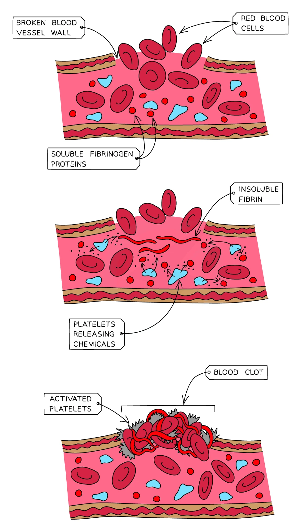
A condition called haemophilia is caused by inheriting a defective version of one of the clotting proteins. People with haemophilia bleed for much longer than normal after even minor cuts, because their clots form very slowly.
Common exam question
Describing the role of platelets in clotting
Question: Describe the role of platelets in the body, or suggest why stored artificial blood, made only of red cells in salt solution, does not clot (1–3 marks).
Set in 2 of the 23 papers. For the 3-mark version give three separate ideas: platelets help the blood to clot (converting soluble fibrinogen into insoluble fibrin is accepted as this point); the clot prevents further blood loss and stops the bleeding; and it prevents pathogens entering the wound, so stops infection. Each is one mark, so a one-idea answer such as "platelets seal wounds" cannot earn all three. When the question asks why stored artificial blood will not clot, the one-mark answer is simply that it has no platelets (no fibrinogen is also allowed).
9. Heart rate and exercise
A typical adult's resting heart rate is around 60 to 80 beats per minute (bpm). The heart rate is set by a special patch of cells in the right atrium called the pacemaker, which sends out regular electrical signals that trigger each heartbeat.
Why heart rate changes with exercise
When you exercise, the muscles need extra energy for every contraction, so the muscle cells step up the rate of respiration. Faster respiration means:
- More oxygen and glucose needed at the muscles
- More carbon dioxide produced that needs removing
- More heat produced that needs distributing or losing
The nervous system responds by speeding up the heart rate, which:
- Pumps more oxygen-rich blood to the muscles per minute
- Removes carbon dioxide from the muscles faster
Each individual heartbeat also pumps more strongly (technically the stroke volume, meaning how much blood is ejected per beat). The combined effect is that cardiac output can rise to roughly five times its resting value during hard exercise.
Adrenaline
The hormone adrenaline, released by the adrenal glands during exercise or fear, also makes the heart beat faster and harder. This is part of the body's fight-or-flight response, preparing the muscles for sudden action.
Heart rate after exercise
The heart rate does not drop straight back to normal when you stop. It stays elevated for several minutes because the body is still paying back the oxygen debt built up during anaerobic respiration in the muscles. The faster the heart rate returns to normal, the fitter the person is, giving a simple way to estimate fitness from heart-rate recovery time.