1. What respiration is
Respiration is the chemical process inside every living cell that releases energy from glucose. The released energy is transferred to ATP, which the cell can then spend on any process that needs energy
Some important things to notice about that definition:
- Respiration happens in every living cell, all the time. Plant cells respire just as much as animal cells, and they do it day and night.
- Respiration is not the same as breathing. Breathing (or gas exchange) is how oxygen gets into the body and carbon dioxide gets out. Respiration is the chemical reaction inside the cells that uses that oxygen to release energy.
- Energy is not produced or made by respiration. Energy is released from the bonds of glucose and transferred to ATP. Exam mark schemes are strict on this point, so never write "respiration produces energy".
What ATP does for the cell
The energy released by respiration is stored in the chemical bonds of a molecule called ATP (adenosine triphosphate). ATP is the "energy currency" of the cell. When ATP is broken down, the stored energy is released right where it is needed. Cells use ATP for:
- Building large molecules from smaller ones (proteins from amino acids, starch from glucose, DNA replication, etc.)
- Muscle contraction for movement
- Active transport across membranes (pumping ions against a concentration gradient)
- Cell division (mitosis and meiosis)
- Generating body heat in warm-blooded animals, to keep enzymes at their optimum temperature
- Nerve impulses (resetting the resting potential after each impulse)
2. Aerobic respiration
uses oxygen. It takes place inside the mitochondria of the cell and represents the complete breakdown of glucose. Aerobic respiration is by far the more efficient form: a single molecule of glucose can be used to make roughly 30 molecules of ATP through the aerobic pathway, compared with only 2 ATP through anaerobic respiration.
Word equation
glucose + oxygen → carbon dioxide + water (+ energy released)
Balanced symbol equation
C₆H₁₂O₆ + 6 O₂ → 6 CO₂ + 6 H₂O
In words: one molecule of glucose reacts with six molecules of oxygen to give six molecules of carbon dioxide and six molecules of water, plus a large amount of energy stored in ATP.
Notice that this equation is the exact reverse of photosynthesis. Photosynthesis builds glucose using light energy; respiration breaks glucose back down to release that energy.
Exam note: If a question asks for the symbol equation, you must give the symbol equation. Writing the word equation when the symbol equation is asked for will lose the mark.
Exam tip
Balance the symbol equation for aerobic respiration
The balanced symbol equation for aerobic respiration has been asked for once, for two marks, and it is cheap to learn: C₆H₁₂O₆ + 6O₂ → 6CO₂ + 6H₂O. The marking has two steps: correct formulae with wrong or missing balancing numbers score one mark, and the fully balanced equation scores two. The word equation scores nothing when the symbol equation is asked for, however neatly it is written. Check the balance by counting atoms on each side before you move on: 6 carbon, 12 hydrogen and 18 oxygen.
3. Anaerobic respiration
releases energy from glucose without using oxygen. It is an incomplete breakdown of glucose, so it produces much less ATP per glucose molecule than aerobic respiration. The waste products are also different from the aerobic ones, and they vary between organisms.
Anaerobic respiration in animals (including human muscle)
In a hard sprint, your leg muscles need so much energy that the blood cannot deliver oxygen fast enough to keep up with aerobic respiration. The muscle cells switch to anaerobic respiration to top up their ATP supply.
Word equation for anaerobic respiration in animals:
glucose → lactic acid (+ a little energy)
Notice that no oxygen is needed, and the waste product is , not carbon dioxide. Glucose is not fully broken down, so most of the energy that was in the glucose ends up locked inside the lactic acid molecules.
Two consequences for the body:
- Lactic acid builds up in the muscle, lowering its pH. Acidic conditions slow down the muscle's enzymes and cause the familiar burning and aching feeling.
- After the exercise, the body must break down the lactic acid using oxygen, in the liver. This takes time and is why you continue to breathe heavily for several minutes after stopping. Your body is still supplying the extra oxygen needed to clear the lactic acid. This extra oxygen demand is called the (or "excess post-exercise oxygen consumption").
Exam tip
Link lactic acid to the oxygen debt
Whenever exercise or recovery appears in a question, write the chain in full: the muscles respired anaerobically, so lactic acid built up, and extra oxygen is needed to break it down: the oxygen debt. In the two-mark question on why breathing stays fast after exercise, those three ideas are separate marking points and any two score. "Breaking down lactic acid" alone earns only the lactic acid mark; "oxygen is needed to break down the lactic acid" earns that mark and the oxygen-debt mark. It scores elsewhere too: a trained athlete has less anaerobic respiration and a smaller oxygen debt.
Anaerobic respiration in yeast and plants (fermentation)
In yeast cells and in some plant cells, anaerobic respiration produces different waste products:
glucose → ethanol + carbon dioxide (+ a little energy)
This special case of anaerobic respiration in yeast is called and is industrially important:
- Brewing: yeast ferments the sugars in fruit or grain into ethanol (alcohol), giving beer, wine and spirits.
- Baking: yeast ferments sugars in bread dough. The carbon dioxide produced makes the dough rise; the small amount of ethanol evaporates during baking.
- Biofuels: ethanol from fermented crops is used as a renewable fuel additive.
Common exam question
Naming the products of anaerobic respiration
Question: Name the gas or the substances that yeast produces by anaerobic respiration, or explain why the acidity of a culture rises as its bacteria respire anaerobically (1–2 marks).
Asked in 5 of the 23 papers. Yeast makes ethanol and carbon dioxide: in the multiple-choice version "ethanol only" is wrong because carbon dioxide is also made, and any row containing lactic acid is wrong because yeast never makes it. Bacteria in milk, like muscle cells short of oxygen, make lactic acid instead (lactate is accepted), and that is what lowers the pH of yoghurt, so the two marks there come from any two of anaerobic respiration, by bacteria, and lactic acid produced.
When a question asks which liquid identifies the gas, name limewater and say it turns cloudy (or hydrogen carbonate indicator, which turns yellow or orange). The mark for the change is only given when the indicator you named is the right one.
Common exam question
Method questions on the yeast respiration practical
Question: Give the purpose of the oil layer, say what apparatus is needed to keep or change the temperature, or describe how to measure the rate of gas production (1–2 marks per part).
Asked in 4 of the 23 papers. The layer of paraffin oil stops oxygen getting in, so the yeast respires anaerobically: say that oxygen is kept out, because "it ensures anaerobic respiration" is ignored. A water bath keeps the temperature constant; to vary or monitor it you also need a thermometer or thermostat, and a timer if a rate is being measured. To measure the rate, collect the gas in a gas syringe or measuring cylinder and record the volume in a set time (or the time to collect a set volume): the second mark needs both volume and time, and counting bubbles earns nothing when the stem already measures volume.
Side-by-side comparison
| Aerobic respiration | Anaerobic respiration | |
|---|---|---|
| Oxygen needed? | Yes | No |
| Glucose breakdown | Complete | Incomplete |
| Where it happens | Mitochondria | Cytoplasm |
| Products (animals) | Carbon dioxide + water | Lactic acid only |
| Products (yeast / plants) | Carbon dioxide + water | Ethanol + carbon dioxide |
| Energy released per glucose | A lot (≈ 30 ATP) | A little (≈ 2 ATP) |
| Industrial use | None directly | Fermentation: brewing, baking, biofuels |
4. Core practical: investigating respiration
Showing that respiration produces carbon dioxide
Principle: living cells release carbon dioxide during respiration. Limewater (calcium hydroxide solution) turns cloudy white when carbon dioxide is bubbled through it, so it can be used to detect the CO₂ given off by a respiring organism.
Apparatus: three sets of boiling tubes, each containing a delivery tube that bubbles into a second boiling tube of limewater. The three test tubes hold:
- Tube A: 10 germinating peas on damp cotton wool (the test)
- Tube B: 10 boiled (dead) peas on damp cotton wool (control for whether the organism is alive)
- Tube C: 10 glass beads on damp cotton wool (control for whether the seed material itself matters)
Method:
- Set up the three identical test tubes as above, each sealed with a bung connected by a delivery tube to a fresh tube of limewater. Make sure each delivery tube dips below the limewater so any gas bubbles through it.
- Leave the whole apparatus in a warm place (25–30 °C) for several hours or overnight.
- Check each limewater tube for cloudiness.
Expected results:
| Tube | What's inside | Limewater result | Why |
|---|---|---|---|
| A | Germinating peas | Cloudy | The seeds are alive and respiring, releasing CO₂ |
| B | Boiled peas | Stays clear | Boiling killed the cells, so they cannot respire |
| C | Glass beads | Stays clear | No living material, no respiration |
The boiled-seed control rules out the possibility that the seed material (rather than living respiration) is releasing CO₂. The glass-bead control rules out the apparatus or air itself being responsible.
Common exam question
Comparing inhaled and exhaled air
Question: Name the solution that compares carbon dioxide in inhaled and exhaled air, explain the change in each tube, or explain why the gas percentages differ (1–3 marks per part).
Asked in 3 of the 23 papers. Name limewater or hydrogen carbonate indicator. Exhaled air goes through the tube that dips into the liquid; you could not inhale through it without sucking liquid up. That tube goes cloudy or milky (yellow with the indicator), while the inhaled-air tube stays clear (stays red or orange), or changes slowly, because exhaled air carries more carbon dioxide, made by respiration. Colours must match the solution named, "changes colour" scores nothing, and "cloudy in both" earns one mark at most. Inhaled air has less carbon dioxide, not none.
For gas percentages: oxygen diffuses into the blood and is used in respiration, carbon dioxide is produced by respiration, and nitrogen is not used. Exhaled air is also warmer and holds more water vapour.
Showing that respiration releases heat
Principle: aerobic respiration releases energy, and not all of that energy is captured as ATP. A noticeable fraction is released as heat, which can be detected with a thermometer in a well-insulated container.
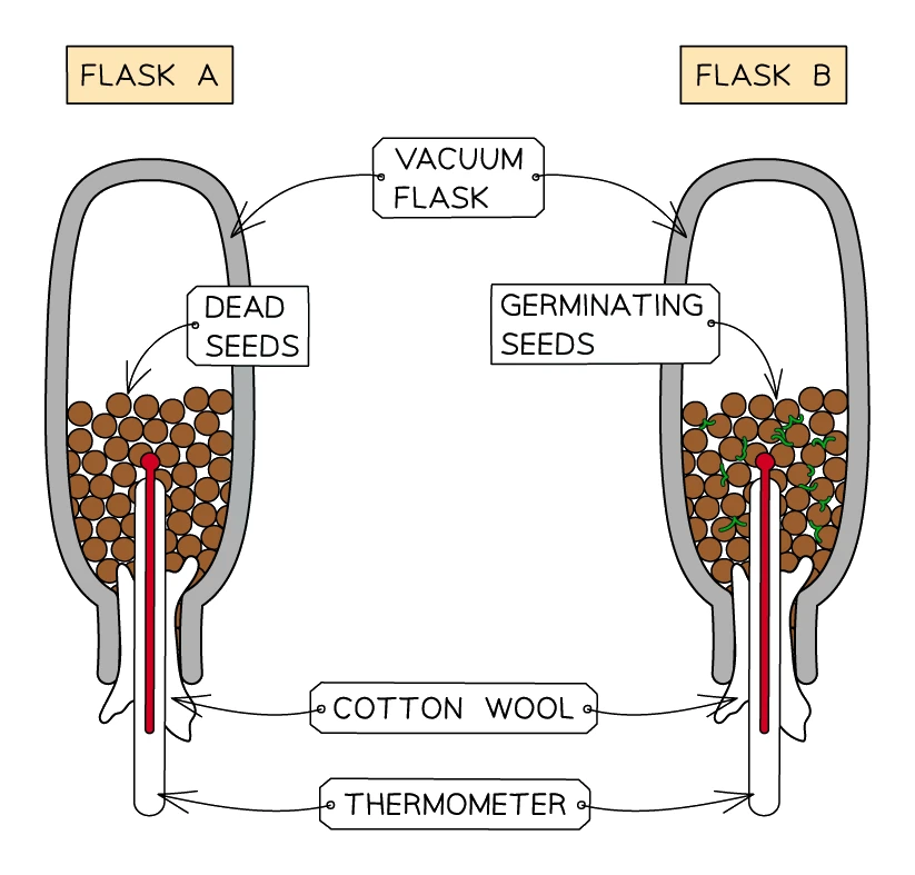
Apparatus: two vacuum flasks, each containing a thermometer and damp cotton wool. The flasks are inverted (mouth-down) so warm air does not simply float out. One flask holds germinating seeds, the other holds boiled (dead) seeds as a control.
Both the seeds and the flasks are first sterilised with weak bleach to kill any microbes. This step is crucial: without it, microbes growing on the seeds will respire themselves and produce heat, giving misleading results.
Method:
- Sterilise both flasks and both sets of seeds with weak bleach.
- Place boiled seeds in flask A and germinating seeds in flask B.
- Insert a thermometer into each flask with the bulb buried in the seeds.
- Plug the mouth of each flask with cotton wool (holds the thermometer in place and reduces heat loss).
- Invert the flasks and leave them in a place with constant room temperature for 2–3 days.
- Record the temperature inside each flask at the start and at the end.
Expected results: the temperature in the flask with germinating seeds rises by several degrees (e.g. 20 °C → 25 °C). The temperature in the flask with dead seeds stays the same as the room.
Common exam question
Explaining the effect of temperature on respiration rate
Question: Explain the effect of increasing the temperature on the rate of gas production by yeast, or on how far the bubble moves in a respirometer (3–4 marks).
Asked in 4 of the 23 papers, and the marks are enzyme marks. Warmer molecules have more kinetic energy and move faster, so enzyme and substrate collide more often and form more enzyme-substrate complexes, so the rate of respiration rises (in a respirometer more oxygen is used up, so the bubble moves further). Give the energy to the molecules, not to the bubble, and do not simply restate that the bubble moves more, which is not credited. Where the range runs past the optimum, or no range is given, add the third step: the active site changes shape, the substrate no longer fits and the enzyme denatures (the enzyme, never "the yeast"). Where the rise stops short of the optimum, the credited idea is that the enzymes are nearer their optimum temperature.
5. Gas exchange: getting oxygen to the cells
Gas exchange and respiration are easy to confuse. To keep them separate:
- Respiration is the chemical reaction inside cells that uses oxygen and produces CO₂ (covered above).
- Gas exchange is the physical movement of oxygen into the body and CO₂ out of the body, by diffusion across an exchange surface.
Why multicellular organisms need a gas exchange surface
A single-celled organism like Amoeba has a huge surface-area-to-volume ratio. Oxygen from the water can simply diffuse through its cell membrane straight into the cytoplasm fast enough to keep every part of the cell supplied. No special apparatus needed.
For a multicellular animal like a human, the inner cells of the body are far too deep for diffusion alone to supply them with oxygen at a useful rate. The body solves this by having:
- A specialised gas exchange surface (the lungs) with a huge surface area and very thin walls
- A transport system (the blood) to carry oxygen from the exchange surface to every cell
Features of a good gas exchange surface
Any good gas exchange surface, in any animal, has the same four adaptations:
- Large surface area so a lot of oxygen can cross at once
- Thin walls (one cell thick where possible) so the diffusion distance is short
- Steep concentration gradient maintained by good ventilation on the outside and good blood supply on the inside
- Moist surface, because the gases must dissolve before they can diffuse through the cells
6. The human respiratory system
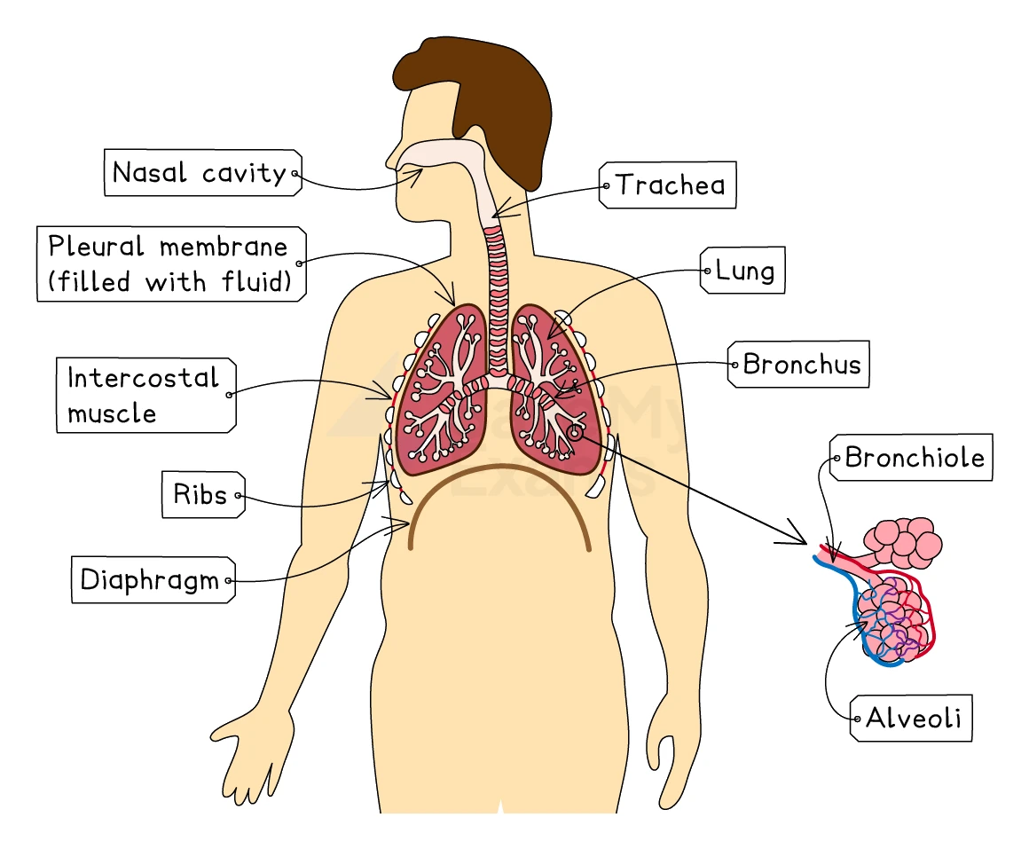
The human gas-exchange organ is the lungs, housed inside the thorax (the chest cavity above the diaphragm). The thorax contains:
| Structure | What it does |
|---|---|
| Ribs | A bony cage that protects the lungs and the heart, and provides anchor points for the muscles that move air in and out |
| Intercostal muscles | Two layers of muscle between each pair of ribs. The external intercostals pull the ribs up and out during inhalation; the internal intercostals pull the ribs down and in during forced exhalation |
| Diaphragm | A thin sheet of muscle below the lungs separating the thorax from the abdomen. It contracts and flattens during inhalation and relaxes back into a dome shape during exhalation |
| Trachea (windpipe) | The single tube carrying air from the throat down towards the lungs. Held open by C-shaped rings of cartilage so it cannot collapse |
| Bronchi (singular: bronchus) | The two large tubes that branch off the trachea, one to each lung |
| Bronchioles | Smaller and smaller tubes that branch out from the bronchi, like the twigs on a tree |
| Alveoli (singular: alveolus) | Tiny balloon-like air sacs at the very end of each bronchiole. This is where gas exchange happens |
| Pleural membranes | Thin double-layered membranes that surround each lung and line the inside of the thorax, with a slippery fluid in between so the lungs can move smoothly against the chest wall |
Order of air flow during inhalation:
mouth/nose → trachea → bronchus → bronchioles → alveoli
Common exam question
Naming the structures air passes through
Question: Complete a passage about the lungs with the missing structures, name a labelled structure on a diagram of the thorax, or pick the order air passes through from the mouth to an alveolus (1 mark each, up to 6 in a passage).
Asked in 4 of the 23 papers. Learn the route as a sequence: trachea (windpipe is accepted), the two bronchi, many bronchioles, then the alveoli, where gas exchange happens. The passage also wants the diaphragm as the muscular sheet that inflates the lungs and the intercostal muscles that move the ribs; "internal" or "external" is neither needed nor penalised there. On a diagram, the bronchus is the wide tube that leaves the trachea into each lung and, like the trachea, has rings of cartilage; the narrow tubes with no cartilage that branch towards the air sacs are bronchioles. The oesophagus is not part of the system: it runs behind the trachea to the stomach.
7. Breathing: how air gets in and out
Muscles can only pull, never push. So breathing relies on two sets of muscles working as antagonistic pairs, plus the diaphragm, to change the volume of the thorax. Because gas pressure depends on volume, changing the volume forces air to flow in or out.
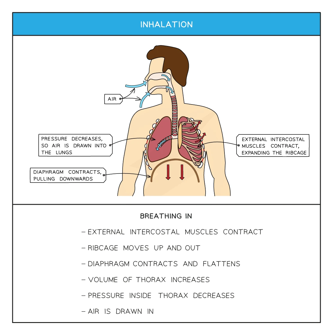
Inhalation (breathing in)
- The external intercostal muscles contract, pulling the rib cage up and out.
- The contracts, flattening from its dome shape downwards.
- Both of these increase the volume of the thorax.
- The pressure inside the lungs falls below atmospheric pressure.
- Air rushes in from the outside, down the trachea, through the bronchi and bronchioles, until it fills the alveoli.
Common exam question
Explaining how breathing in happens
Question: Describe how the diaphragm and intercostal muscles bring about inhalation, in the body or in a bell-jar model of breathing (1–4 marks).
Asked in 4 of the 23 papers; every step is a separate mark, so write them all: the diaphragm contracts and flattens (moves down); the external intercostal muscles contract, raising the rib cage up and out; the volume of the thorax increases; the pressure inside it falls; so air is drawn in. Contracts and flattens are two separate marks, so give both. It is the volume of the thorax, not of the lungs (ignored), and it increases rather than making "more space" (ignored). The diaphragm moves down because it contracts: "relaxes and moves down" is wrong.
In the bell-jar model the rubber sheet is the diaphragm; pulling it down raises the volume and lowers the pressure. Its limits: the rigid jar cannot move like the rib cage, and it has no ribs, intercostal muscles or pleural membranes.
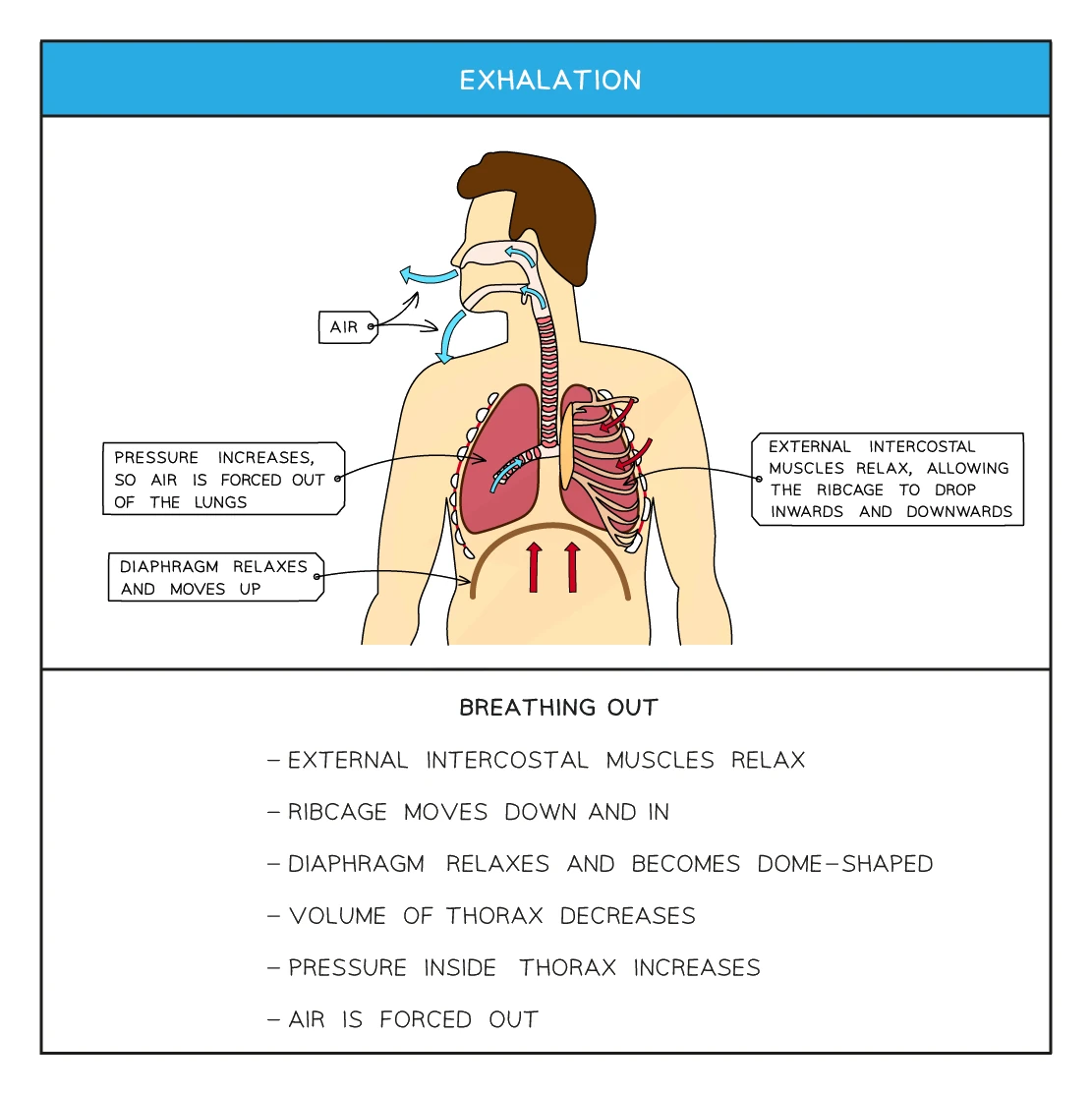
Normal exhalation (breathing out)
- The external intercostal muscles relax, so the rib cage drops down and inwards under its own weight.
- The relaxes, returning to its dome shape pushing up into the thorax.
- The volume of the thorax decreases.
- The pressure inside the lungs rises above atmospheric pressure.
- Air is pushed out of the lungs along the same path in reverse.
Normal exhalation is passive, relying on the elastic recoil of the lungs and rib cage. The internal intercostal muscles do not contract during normal breathing.
Forced exhalation (during exercise)
During hard exercise, the body needs to clear CO₂ much faster. The internal intercostal muscles contract, actively pulling the rib cage further down and in. This reduces the thorax volume even more, forcing air out faster. The two intercostal muscle layers are working as antagonistic pairs: when one contracts the other relaxes.
| Stage | External intercostals | Diaphragm | Thorax volume | Air pressure in lungs | Air flow |
|---|---|---|---|---|---|
| Inhalation | Contract | Contract (flattens) | Increases | Decreases | In |
| Normal exhalation | Relax | Relaxes (domes up) | Decreases | Increases | Out |
| Forced exhalation | Relax (internal intercostals contract) | Relaxes more | Decreases more | Increases more | Out faster |
8. The alveoli: where gas exchange happens
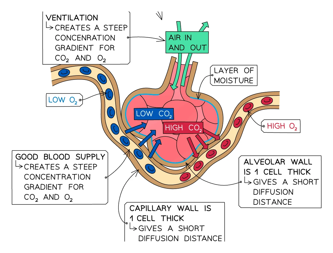
Each lung contains roughly 300 million , the tiny air sacs at the very end of each bronchiole. The alveoli are where oxygen actually crosses from the air into the blood, and where CO₂ goes the other way.
How the alveoli are adapted for gas exchange
Each adaptation matches one of the four general features of a good exchange surface:
- Large surface area: there are hundreds of millions of alveoli per lung. If you flattened all the alveolar walls of one human's lungs into a single sheet, the area would be around 70 m², about half a tennis court.
- Thin walls: each alveolus is just one cell thick, and so are the capillaries wrapped around it. The total diffusion distance from air to blood is therefore only about 0.5 μm.
- Steep concentration gradient: maintained by two things working together. The lungs are constantly ventilated with fresh air (rich in oxygen, low in CO₂), and the capillaries are constantly carrying blood away (taking the absorbed oxygen with it, replacing it with deoxygenated blood).
- Moist surface: each alveolus is lined with a thin film of fluid. Oxygen and CO₂ both dissolve in this fluid before crossing the cell wall.
Exam tip
Pair each alveolus adaptation with its reason
The four-mark question on how a single alveolus is adapted is marked in pairs, a structure and its function, and the function mark is not given without its structure. Safe pairs: the wall is one cell thick, so the diffusion path is short; a rich blood supply of capillaries maintains a steep concentration gradient; a moist lining lets gases dissolve and pass through the wall. Do not spend a pair on surface area: for a single alveolus it is ignored. Gas exchange happens only at the alveoli, not in the trachea, bronchi or bronchioles.
Direction of diffusion
- Oxygen moves into the blood, from a high concentration in the alveolar air to a lower concentration in the deoxygenated blood arriving in the capillaries.
- Carbon dioxide moves out of the blood, from a high concentration in the arriving blood to a lower concentration in the alveolar air, then is breathed out.
9. Exercise and breathing
During exercise the muscles use far more ATP than at rest, so they need far more glucose and oxygen and produce far more CO₂. The body responds in several ways:
- Breathing rate increases, with more breaths per minute drawing in fresh oxygen faster.
- Depth of breathing increases, so each breath moves a larger volume of air (the "tidal volume" rises).
- Heart rate increases to pump the oxygen-rich blood to the muscles faster.
The combined effect is that more oxygen reaches the muscles per minute and more CO₂ is cleared per minute.
Common exam question
Explaining the change in breathing rate during exercise
Question: Explain the change in breathing rate during exercise, or comment on the effect of exercise on ventilation rate using the data given (2–4 marks).
Asked in 3 of the 23 papers. Describe the data first, with numbers: the rate rises, then reaches a maximum or levels off (saying it "slows down" is ignored, and a description mark needs the time or speed at which the rise stops). Then explain: more oxygen is taken in and more carbon dioxide is released; the oxygen reaches the muscles for respiration, releasing more energy as ATP for faster muscle contraction. The depth of breathing (volume per breath) rises as well: at low effort the extra air comes from deeper breaths alone, and as the depth levels off the rate takes over. The plateau is because the chest cannot inflate and deflate any faster. Each of these is a separate mark, so give several short points rather than one long sentence.
Oxygen debt
If the exercise is so intense that even maximal breathing cannot keep up with the muscles' oxygen demand, the muscles switch to and produce . After the exercise is over, the body needs extra oxygen to break the lactic acid back down. This is the (sometimes called excess post-exercise oxygen consumption, EPOC).
You "pay back" the oxygen debt by continuing to breathe hard for several minutes after stopping, even though the muscles are no longer working. The extra oxygen is being used by your liver to convert lactic acid back into harmless products (glucose, CO₂ and water).
Investigating the effect of exercise on breathing rate
A simple practical to demonstrate this:
- Have each volunteer sit quietly for 1 minute, then count their number of breaths per minute at rest.
- The volunteer then performs a controlled exercise (e.g. running on the spot, or step-ups at a set pace) for 1 minute.
- Immediately after exercise, count the breaths per minute again.
- Calculate the change in breathing rate: change = breathing rate after − breathing rate before.
- Repeat with several volunteers and take a mean to reduce variation.
Expected result: breathing rate is markedly higher after exercise. The increase ranges from a few breaths per minute (for light exercise) to more than double the resting rate (for vigorous exercise).
Limitations:
- Different people have different fitness levels. Control this by using a similar age and fitness range, or report results for each person separately.
- Exercise intensity is hard to standardise. Use a fixed exercise (step-ups at a metronome pace) rather than free running.
- Counting breaths immediately after exercise is fiddly. Count for 15 seconds then multiply by 4 to catch the result before the rate falls back.
Common exam question
Describing an investigation into exercise and breathing
Question: Describe a method to show the effect of exercise on breathing rate, or a simple experiment to show that exercise increases the carbon dioxide a person breathes out (3 marks).
Asked in 2 of the 23 papers. For breathing rate: count the breaths per minute at rest, then again during or straight after a set exercise; a spirometer is accepted for either count, but measuring heart rate instead loses both of those marks. Then repeat with more people and control age, sex, fitness and the amount or duration of exercise. For carbon dioxide: breathe out through limewater (or hydrogen carbonate indicator) and time how long it takes to go cloudy (or change colour) before and after exercise, keeping the volume of limewater, the room temperature and the person the same, then repeat. When a data question asks how to make the investigation more reliable, the one-mark answer is to repeat it, or use more people, and calculate a mean.
10. The effects of smoking on the gas exchange system
Cigarette smoke contains several chemicals that damage the gas exchange system and the rest of the body.
Three of the key chemicals
| Chemical | Where it acts | What it does |
|---|---|---|
| Nicotine | Heart and blood vessels | Highly addictive. Raises heart rate and constricts blood vessels, raising blood pressure. The higher pressure damages artery walls and increases the risk of clots, heart attack and stroke |
| Carbon monoxide | Red blood cells | Binds irreversibly to haemoglobin, taking the place where oxygen would have bound. Reduces the blood's oxygen-carrying capacity, forcing the heart and lungs to work harder to deliver the same oxygen |
| Tar | Lining of the airways | A carcinogen (cancer-causing chemical). Builds up in the airways, irritates the lining, and causes chronic bronchitis, emphysema and lung cancer |
Three smoking-related lung diseases
Chronic bronchitis
- Tar inflames the lining of the airways. In response, the goblet cells and mucus glands grow larger and pour out far more mucus than they would in a healthy lung.
- The mucus builds up in the smallest bronchioles, blocking them and creating an ideal place for bacteria to grow. Frequent chest infections follow.
- Tar also damages the cilia on the airway lining, so the natural mechanism that sweeps mucus up out of the lungs no longer works.
- The smoker has to cough to clear the mucus, which is the familiar "smoker's cough".
- Repeated infections attract phagocytes (a type of white blood cell) into the lungs. The phagocytes release an enzyme called elastase, which destroys the elastic fibres that normally hold the alveolar walls together.
- Without elastic fibres, the alveoli cannot recoil properly. Many alveoli burst, merging into larger air spaces.
- The total surface area for gas exchange falls, sometimes to a tenth of the normal value.
- The patient becomes breathless and wheezy with even gentle activity, and in late stages may need a constant oxygen supply.
Lung cancer
- Many of the chemicals in tar are carcinogens that can damage the DNA of cells lining the airways.
- A damaged cell can become a cancer cell that divides uncontrollably, forming a tumour.
- Smoking is the single biggest cause of lung cancer; the risk falls steeply if the smoker quits.
Common exam question
Describing the effects of smoking on the lungs and blood
Question: Describe what smoking does to the lungs, or how carbon monoxide affects the body (2–5 marks).
Asked in 2 of the 23 papers. The five-mark lungs question is marked one point per named effect, so name every step: smoke destroys the cilia, so mucus builds up and cannot be removed, leading to infection; the alveoli are damaged and lose surface area (emphysema); the airways become inflamed and narrowed (bronchitis), together called COPD; and the carcinogens cause lung cancer. For the blood: carbon monoxide binds to haemoglobin, forming carboxyhaemoglobin, so less oxygen is carried and cells respire less (or more anaerobically) and release less energy; "forms carboxyhaemoglobin rather than oxyhaemoglobin" earns both marks of the two-mark version. The same chain earns marks when a data question asks about smoking in pregnancy.
Coronary heart disease
Smoking is also a major risk factor for coronary heart disease. Nicotine raises blood pressure, carbon monoxide reduces oxygen delivery, and both put strain on the coronary arteries that supply the heart muscle. Fatty deposits called atheromas are more likely to build up inside these arteries, eventually leading to a heart attack if a coronary artery becomes blocked.