1. The biological molecules in food
Almost everything in your food (and in your own body) is built from three families of large biological molecules:
| Molecule | Elements it contains | Smaller building blocks |
|---|---|---|
| Carbohydrates | Carbon, hydrogen, oxygen | Simple sugars (e.g. glucose) |
| Proteins | Carbon, hydrogen, oxygen, nitrogen (and sometimes sulfur) | Amino acids |
| Lipids (fats and oils) | Carbon, hydrogen, oxygen | Glycerol and fatty acids |
All three are organic molecules because they are based on carbon. Proteins are the only one of the three that always contains nitrogen, which is why plants need nitrate ions from the soil to build them.
Carbohydrates
- Glucose (C₆H₁₂O₆) is a single simple sugar molecule.
- Maltose is made when two glucose molecules join together.
- , glycogen and cellulose are all very long chains made from many glucose molecules joined together.
- Starch is how plants store glucose (in roots, tubers, seeds)
- Glycogen is how animals store glucose (in the liver and in muscles)
- Cellulose is what plant cell walls are made of
- These long chains are insoluble in water, which is why they make good storage molecules: they stay where the cell puts them and do not affect the cell's water balance.
Proteins
- Proteins are long chains of amino acids joined together.
- There are about 20 different amino acids, and they can be arranged in any order. A different sequence makes a different protein, which is why there are hundreds of thousands of different proteins doing different jobs in the body.
- The amino acid sequence determines the shape of the protein. The shape, in turn, determines what the protein does. Examples:
- (catalysts that speed up chemical reactions)
- Haemoglobin (the oxygen-carrying protein in red blood cells)
- Keratin (the protein in hair, nails and skin)
- Antibodies (used by the immune system)
Lipids
- Lipids are built from glycerol and fatty acids. One glycerol plus three fatty acids gives a single triglyceride, the most common lipid in food.
- Solid lipids at room temperature are called fats (butter, lard).
- Liquid lipids at room temperature are called oils (olive oil, sunflower oil).
2. A balanced diet
A balanced diet contains all of the necessary nutrient groups in the right proportions. The seven essential groups are:
| Food group | Function in the body | Common food sources |
|---|---|---|
| Carbohydrates | Main source of energy | Bread, rice, pasta, potatoes, cereals |
| Proteins | Growth and repair of tissues; making enzymes, antibodies, hormones | Meat, fish, eggs, beans, nuts, dairy |
| Lipids | Long-term energy storage and insulation; part of every cell membrane | Butter, oils, nuts, oily fish |
| Dietary fibre | Provides roughage so the muscles of the gut can push food along | Vegetables, fruit, wholegrain bread, beans |
| Vitamins | Needed in tiny amounts to keep the body working | Fruit, vegetables, dairy, meat |
| Minerals | Needed in tiny amounts for specific roles (e.g. calcium for bones, iron for blood) | Dairy, meat, leafy green vegetables |
| Water | Solvent for chemical reactions; transport medium in blood; lost as sweat and urine | Drinks, fruit, vegetables |
Key vitamins and minerals to know
- Vitamin A is used to make the pigment in the retina that lets you see in dim light. A deficiency causes night blindness. Found in liver, dairy, leafy green vegetables and orange-yellow vegetables.
- Vitamin C is needed to make collagen, the protein that holds skin, gums and connective tissues together. Severe deficiency causes scurvy (bleeding gums, slow wound healing). Found in citrus fruits, peppers and green vegetables.
- Vitamin D helps the body absorb calcium from food and is needed for strong bones and teeth. Severe childhood deficiency causes rickets (soft, bowed bones). Found in oily fish and dairy products, and made by the skin in sunlight.
- Calcium is needed for bones and teeth, and helps blood clot at a wound. A long-term deficiency leads to osteoporosis (weak, brittle bones). Found in milk, cheese and yoghurt.
- Iron is needed to make haemoglobin in red blood cells. A deficiency causes anaemia (tiredness, pale skin, breathlessness) because the blood cannot carry enough oxygen. Found in red meat, liver, leafy green vegetables.
Why energy needs vary
Different people need different amounts of energy depending on:
- Age: children and teenagers need a lot of energy for growth. An adult's energy needs slowly fall after middle age.
- Activity level: a person who exercises a lot or does manual work respires faster, so they need more energy.
- Sex: on average, men have more muscle and so use more energy than women of the same mass.
- Pregnancy and breastfeeding: a pregnant woman needs extra energy, protein, calcium and iron to build the fetus's body and her own larger blood supply. A breastfeeding mother needs extra energy and calcium to produce milk.
An unbalanced diet causes malnutrition. Too little of one group causes a deficiency disease (e.g. scurvy, rickets, anaemia). Too much energy intake versus output causes obesity, which raises the risk of heart disease, type 2 diabetes and some cancers.
Common exam question
Comparing diets from a nutrition table
Question: Use the table and your own knowledge to discuss which food is more suitable for a named person, or the long-term effects of a diet that differs from the recommended daily amounts (4–5 marks).
Asked in 6 of the 23 papers. Most schemes pay for a comparison tied to its consequence for that person: more energy, fat or sugar is stored as fat, so weight gain and a higher risk of diabetes and heart disease; enough protein means normal growth and repair; too little calcium risks rickets or osteoporosis, too little vitamin C scurvy, too little iron anaemia, too little fibre constipation. Two schemes cap a difference with no consequence at one mark, and one caps at three the marks for nutrient functions with no link to a shortage or excess. You may argue from either food (the converse is allowed), and a closing judgement earns a mark, including that weight depends on the whole diet and on activity.
Worked example
Mass of food that supplies a daily amount of a nutrient
The label on a 150 g pot of yoghurt states that it contains 180 mg of calcium. A child needs 900 mg of calcium each day. Calculate the mass of this yoghurt that would supply the child's daily calcium.
Solution:
- Calcium in 1 g of yoghurt: 180 ÷ 150 = 1.2 mg
- Mass of yoghurt needed: 900 ÷ 1.2 = 750 g
- Or scale up directly: 900 ÷ 180 = 5, so 5 × 150 = 750 g
Either route scores, and a correct answer with no working earns both marks. Two schemes give the method mark for the per-gram value or the scale factor; the third asks for two significant figures and gives an unrounded answer only one of the two marks, so round at the end, to the precision asked.
3. Food tests
Four standard food tests detect the four major nutrient groups. Each works by adding a reagent to a food sample and watching for a specific colour change.
Preparing a food sample
For solid food, before testing:
- Grind the food into a paste using a pestle and mortar
- Add a small volume of distilled water and stir
- Filter through filter paper to give a clear solution to test
The four tests
| Test for | Reagent | Method | Positive result |
|---|---|---|---|
| Glucose (reducing sugar) | Benedict's solution | Add a few drops; heat in a water bath for 5 minutes | Blue → brick-red (orange/yellow if less glucose) |
| Starch | Iodine solution | Add a few drops at room temperature | Yellow-brown → blue-black |
| Protein | Biuret solution | Add a few drops at room temperature | Blue → violet (lilac/purple) |
| Lipids | Ethanol (then water) | Mix the sample with ethanol, then add an equal volume of cold water | Clear → cloudy white emulsion |
Notes for each test
- The Benedict's test needs heat. Use a water bath rather than a Bunsen burner directly because the test tubes are easy to break and the Bunsen flame is hard to control. Switch the burner off once the water is hot.
- The iodine test is quick and simple, with no heating needed.
- Biuret solution contains copper sulfate and sodium hydroxide, both of which are irritants. Always wear goggles.
- Ethanol is highly flammable. Keep it well away from any naked flame.
Common exam question
Describing a food test
Question: Describe how to test a sample of food, or of urine, for protein, glucose, starch or lipid (2 marks).
Asked in 9 of the 23 papers, protein and glucose most often. The two marks are the reagent and the positive colour. Protein: add Biuret solution (or sodium hydroxide and copper sulfate), which turns lilac, purple or pink. Glucose, a reducing sugar: add Benedict's solution and heat, giving a red, orange, yellow or green colour; two of the three schemes make the heating a mark of its own, so never leave it out. Starch: add iodine solution, which turns blue-black; black or dark blue is accepted but "purple" is ignored. Lipid: shake the sample with ethanol, then add water, and a cloudy white emulsion forms; Sudan III or the translucent spot on paper are credited alternatives. No scheme gives a mark for the starting colour, so do not spend words on it.
4. Enzymes
What an enzyme is
An enzyme is a that acts as a biological catalyst: it speeds up a specific chemical reaction without being changed or used up itself
Enzymes are essential for life. Without digestive enzymes, breaking down a single meal would take 2 to 3 weeks; with them, it takes around 4 hours. Every metabolic reaction in every living cell is catalysed by an enzyme.
How an enzyme works (the lock-and-key model)
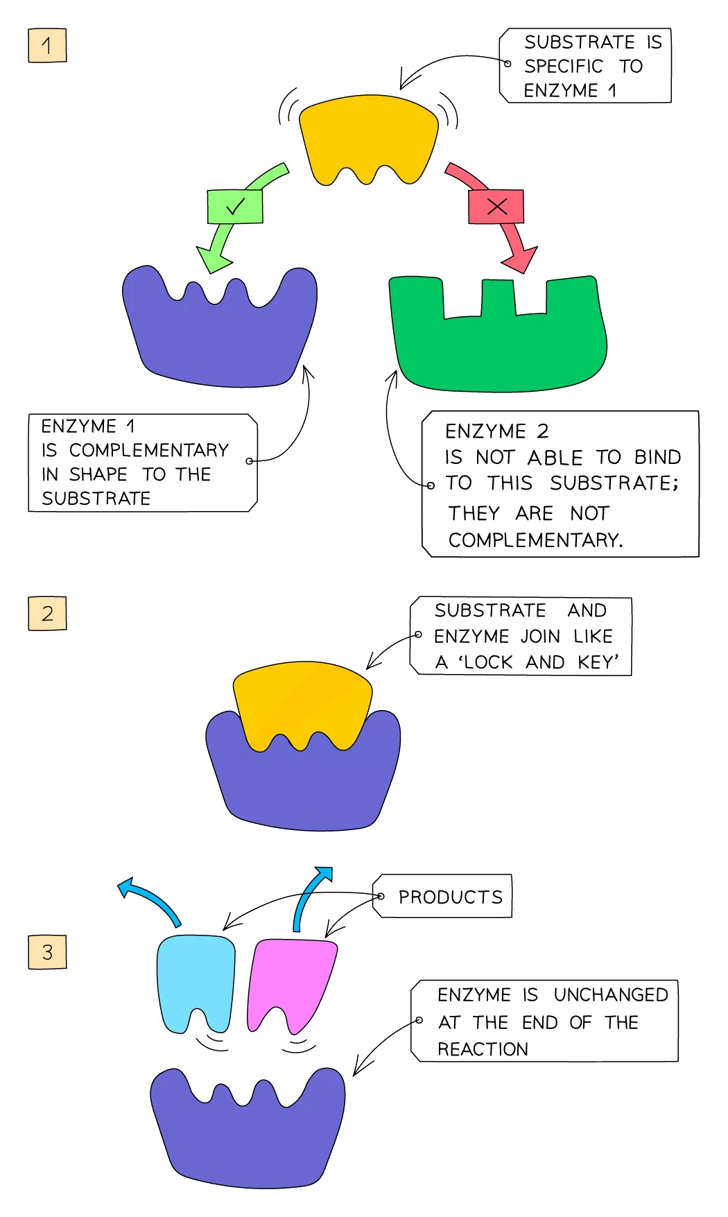
- Each enzyme has a specifically shaped pocket on its surface called the .
- The molecule the enzyme acts on is called the substrate.
- The substrate fits into the active site exactly (the way a key fits into a lock) because the two shapes are complementary.
- The three steps are:
- Substrate and collide randomly in solution
- The substrate enters the active site, forming an enzyme-substrate complex, and the reaction takes place
- The product leaves the active site, which is now free to bind another substrate
Each enzyme is specific to one substrate (or to one type of bond). Amylase only works on starch, for example, because no other molecule has the right shape to fit the amylase active site.
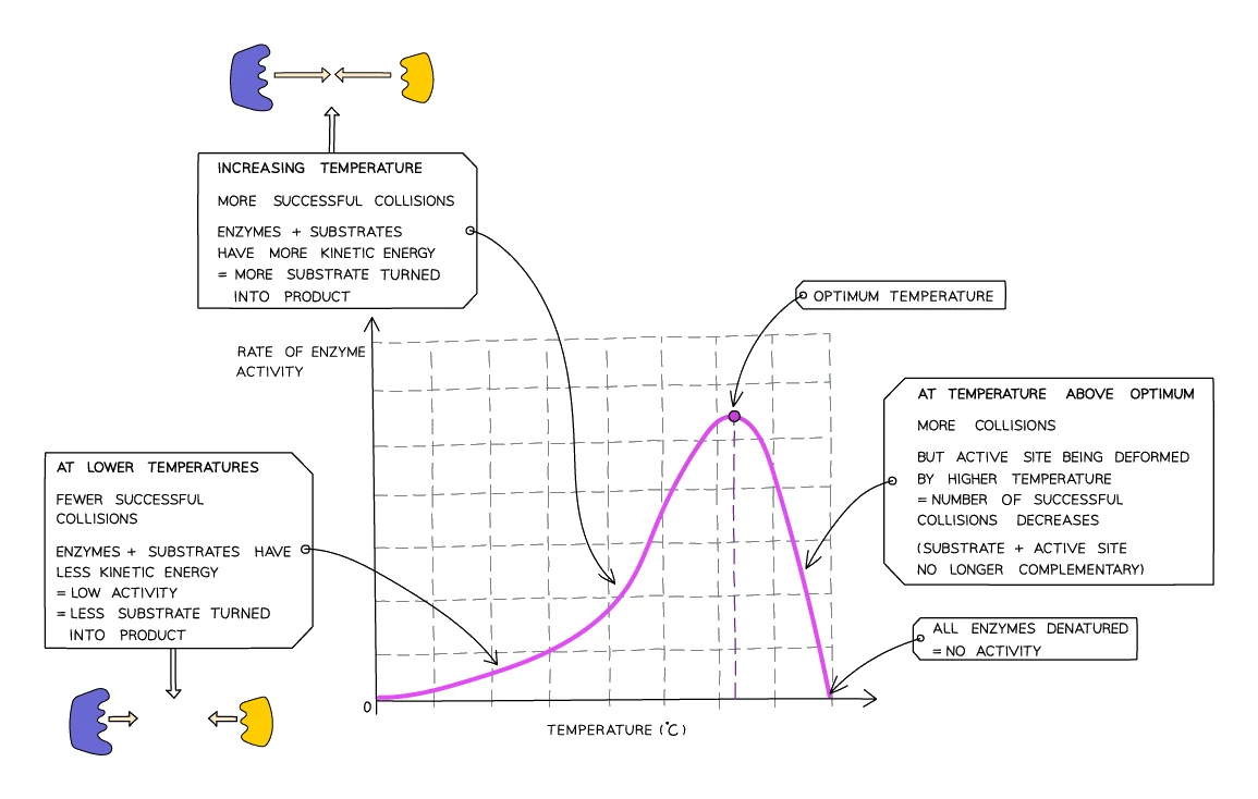
Effect of temperature on enzyme activity
- At low temperatures, enzymes work slowly: the particles have little kinetic energy, so collisions between enzyme and substrate are rare. The reaction is slow but the enzymes are not damaged.
- As temperature rises, collisions happen more often and with more energy. The rate of reaction climbs.
- At the optimum temperature, the enzyme is working as fast as it can. For most human enzymes the optimum is about 37 °C, conveniently the body's normal temperature.
- Above the optimum, the bonds that hold the enzyme's shape together start to break. The active site changes shape and the substrate no longer fits. The enzyme is .
- Denaturation is permanent: cooling the enzyme back down does not restore its shape or its activity.
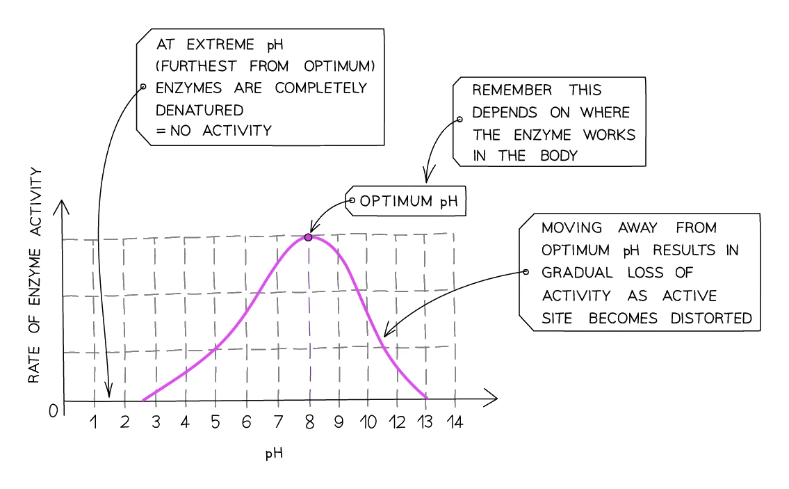
Effect of pH on enzyme activity
- Each enzyme has an optimum pH at which it works fastest.
- Most enzymes have an optimum near pH 7 (neutral).
- Some enzymes have evolved to work in unusual environments:
- Pepsin, the protease in the stomach, has an optimum of pH 2 (very acidic, matching the stomach's hydrochloric acid).
- Enzymes secreted into the small intestine (pancreatic and intestinal enzymes) have an optimum near pH 8 (slightly alkaline).
- Above or below the optimum, the bonds holding the enzyme together break and the active site changes shape: the enzyme denatures.
Common exam question
Explaining the effect of temperature or pH on an enzyme
Question: Explain the effect of increasing the temperature, or of changing the pH, on the rate of an enzyme-controlled reaction, or explain why the reaction had stopped at a high temperature (2–4 marks).
Asked in 12 of the 23 papers, about a digestive enzyme or catalase, or inside a question on yeast respiration or photosynthesis, where the same enzyme points score. Up to the optimum, write the chain: molecules gain kinetic energy and move faster, so more collisions and more enzyme–substrate complexes, so a faster rate (or a shorter time). Above it, bonds break and the enzyme denatures: the active site changes shape and the substrate no longer fits. For pH the same ideas score, and three of the four schemes also give the optimum pH a mark of its own; one scheme gives nothing for its first point unless the word enzyme appears. The enzyme denatures, never the yeast or the cell, and in a question about acid do not blame temperature: that was rejected.
Core practical: temperature and enzyme activity
The effect of temperature on amylase (a starch-digesting enzyme) can be measured using iodine to test for starch:
- Add 5 cm³ of starch solution to a test tube
- Place a drop of iodine in each well of a spotting tile
- Place the starch tube in a water bath at the test temperature (e.g. 20 °C) for 10 minutes to let the solution reach the bath's temperature
- Add 2 cm³ of amylase to the starch and start the stopwatch
- Every minute, transfer a drop of the mixture to a fresh iodine well. The drop turns blue-black if starch is still present, yellow-brown once it has all been digested into maltose
- Record the time it takes for the iodine to stop turning blue-black
- Repeat at a range of temperatures (e.g. 10, 20, 30, 40, 50, 60 °C)
A short time means fast digestion (high enzyme activity). Plotting rate (1 ÷ time) against temperature gives the classic peaked curve, with the optimum around 37 °C for amylase.
Core practical: pH and enzyme activity
The same experiment can be repeated at fixed temperature (in a water bath at 37 °C), using buffer solutions at different pH values (e.g. pH 3, 5, 7, 9, 11) instead of varying temperature. Amylase's optimum pH is around 7.
Common exam question
Method questions on an enzyme investigation
Question: State why the enzyme and substrate are left in the water bath before mixing, give the dependent variable, or name variables that should be controlled in an enzyme investigation (1 mark each).
Set in 4 of the 23 papers. The tubes stand in the bath so that both solutions reach the temperature being tested; "to reach the optimum temperature" is ignored, because each run must start at its own set temperature. The dependent variable is what you measure: the time for the indicator or iodine colour to change, or the volume of gas collected in a set time. For control variables name a quantity, never an "amount", which is ignored: the volume and concentration of enzyme and of substrate, the volume of buffer, the time allowed, the temperature when pH is varied, and the surface area of the substrate. Cutting potato into discs is credited as a larger surface area, so more enzyme meets substrate.
5. The human alimentary canal
What the digestive system does
The digestive system is an organ system whose job is to convert the large insoluble nutrients in food (starch, proteins, lipids) into much smaller soluble units (glucose, amino acids, fatty acids and glycerol). Once small enough, these units pass through the gut wall into the blood and are carried to every cell in the body.
Two kinds of digestion go on:
- Mechanical digestion physically breaks food into smaller pieces. Examples: teeth chewing, stomach churning, bile emulsifying fat. The resulting smaller pieces have more exposed surface, which gives enzymes more places to bind.
- Chemical digestion breaks the bonds that hold large food molecules together, using . This produces the small soluble molecules that can be absorbed.
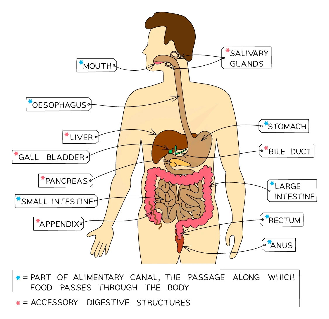
The alimentary canal is the long tube through which food passes from mouth to anus. The accessory organs (liver, gall bladder, pancreas, salivary glands) make digestive juices but food does not pass through them.
Organs of the alimentary canal
| Organ | What happens there |
|---|---|
| Mouth and salivary glands | Teeth break the food into smaller pieces (mechanical digestion). Saliva contains the enzyme amylase, which starts to digest starch into maltose (chemical digestion). The food is rolled into a soft ball called a bolus and swallowed |
| Oesophagus | The tube from mouth to stomach. Waves of muscle contraction (peristalsis) push the bolus down to the stomach |
| Stomach | A muscular bag that churns food into a creamy paste (chyme). The stomach lining secretes hydrochloric acid (pH ~2), which kills bacteria and provides the optimum pH for the stomach's protease enzyme pepsin, which starts to digest proteins |
| Small intestine (duodenum then ileum) | The duodenum receives bile from the gall bladder and enzymes from the pancreas. Digestion of all three nutrient groups is completed here. The ileum is where the small soluble molecules are absorbed into the blood. The pH is slightly alkaline (~pH 8) |
| Large intestine (colon then rectum) | The colon absorbs most of the remaining water from the indigestible material. The rectum stores the resulting faeces until they leave the body through the anus |
| Salivary glands (accessory) | Produce saliva containing amylase and mucus |
| Liver (accessory) | Produces bile, which emulsifies fats and neutralises stomach acid. Also breaks down excess amino acids in a process called deamination, producing urea |
| Gall bladder (accessory) | Stores bile from the liver and releases it into the duodenum |
| Pancreas (accessory) | Makes amylase, protease and lipase (the full set of digestive enzymes) in an alkaline juice, which is squirted into the duodenum |
Common exam question
Identifying organs on a diagram of the gut
Question: Name the parts labelled on a diagram of the alimentary canal, or pick out the oesophagus, the stomach, the part of the large intestine or the structure that contains villi (1 mark each).
Asked in 3 of the 23 papers, once as four written labels on a rabbit's gut and otherwise as multiple choice, on a dog's gut and on a human one. In order the canal is the oesophagus (gullet is accepted), the stomach, the small intestine (ileum, duodenum or jejunum all score) and the large intestine (or colon). Villi are in the small intestine, so choose the long coiled tube, not the wider colon. The liver and pancreas sit beside the canal and food never passes through them. Two papers also ask which organs make amylase, once from the diagram: the salivary glands and the pancreas; the stomach and the colon do not.
6. Peristalsis
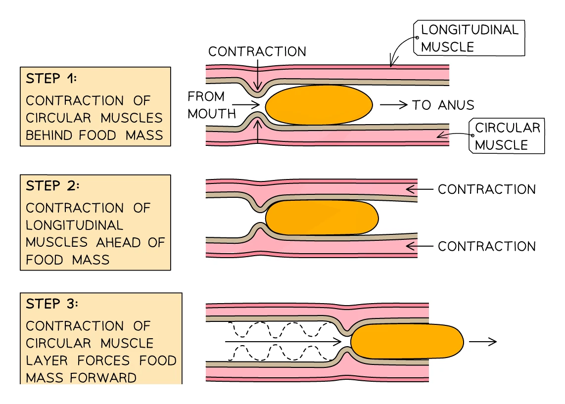
Peristalsis is the wave of muscle contraction that pushes food along the alimentary canal. The wall of the canal contains two layers of muscle:
- Circular muscles run around the canal. When they contract, the diameter of the tube narrows.
- Longitudinal muscles run along the length of the canal. When they contract, the section of tube shortens.
These two layers contract in turn, creating a wave that travels along the canal and pushes the bolus forward, exactly like squeezing toothpaste out of a tube. Peristalsis works against gravity too, which is why an astronaut in space can still swallow food without it floating back up.
Dietary fibre provides the bulk that the muscles can push against. Without enough fibre, peristalsis is sluggish and constipation can develop.
Common exam question
Describing how food is moved along the gut
Question: Describe how food is moved along the gut, or how it travels from the mouth to the stomach after swallowing (2 marks).
Set in 2 of the 23 papers. Name the process, peristalsis, for one mark, then say what happens for the other: the muscles in the wall contract behind the food and push or squeeze it along, through the oesophagus when the question is about swallowing. "The muscles move" is ignored; it is their contraction that scores. The fullest answer has the circular muscles contracting behind the bolus while the longitudinal muscles contract ahead of it.
7. Digestive enzymes
Digestive enzymes fall into three groups, each one catalysing the breakdown of a different nutrient:
| Enzyme group | What it digests | Into | Where it is made |
|---|---|---|---|
| Carbohydrases (amylase, maltase) | Carbohydrates | Simple sugars (e.g. glucose) | Salivary glands, pancreas, small intestine |
| Proteases (pepsin, trypsin) | Proteins | Amino acids | Stomach, pancreas, small intestine |
| Lipases | Lipids | Fatty acids and glycerol | Pancreas, small intestine |
Carbohydrase chain: starch → glucose
Starch is broken down in two steps:
- Amylase (in saliva, then in pancreatic juice in the duodenum) breaks into maltose (a disaccharide made of two glucose molecules).
- Maltase (made by the small intestine) then breaks maltose into individual glucose molecules, which can be absorbed.
Note: the amylase in saliva gets denatured as soon as it enters the acidic stomach (pH 2 destroys its shape), so most starch digestion actually happens in the duodenum after pancreatic amylase is added.
Proteases
- Pepsin in the stomach (optimum pH 2) breaks proteins into shorter chains called polypeptides.
- Trypsin and other proteases from the pancreas and small intestine (optimum pH 8) then finish the job, breaking polypeptides into individual amino acids.
Lipases
Lipase from the pancreas works in the small intestine, breaking into fatty acids and glycerol. Lipase needs the lipids to be in tiny droplets first, which is the job of bile (next section).
Common exam question
Matching digestive enzymes to substrates and products
Question: Complete a table of digestive enzymes, substrates and products, state what starch or protein is digested into, or describe how protein is digested in the alimentary canal (1–4 marks).
Asked in 7 of the 23 papers. One mark per row: amylase digests starch to maltose; maltase digests maltose to glucose; protease digests protein (or polypeptides) to amino acids, with peptides accepted; lipase digests lipids to fatty acids and glycerol. Amylase makes maltose, not glucose. The schemes list fatty acids and glycerol as alternatives, so either earns the lipase row, but write both. Describing protein digestion for 3 marks wants the enzyme (protease, pepsin or trypsin), where it acts (stomach, then duodenum, from the pancreas) and the product (amino acids); absorption is ignored. The same facts answer harder settings: a blocked pancreatic duct means less digestion of all three food groups, and insulin is injected because, being a protein, it would be digested by protease.
8. Bile
What bile is and where it comes from
Bile is a green-yellow alkaline fluid produced by cells in the liver, stored in the gall bladder, and released into the duodenum when food arrives. Bile is not an enzyme (it does not break any chemical bonds), but it has two essential roles in digestion.
Role 1: Neutralising stomach acid
Food leaves the stomach very acidic (pH ~2). The digestive enzymes secreted into the small intestine have their optimum at pH 8 and would be denatured by stomach acid. Bile is alkaline and neutralises the stomach acid as it enters the duodenum, raising the pH to the right level for the small intestine's enzymes to work.
Role 2: Emulsifying lipids
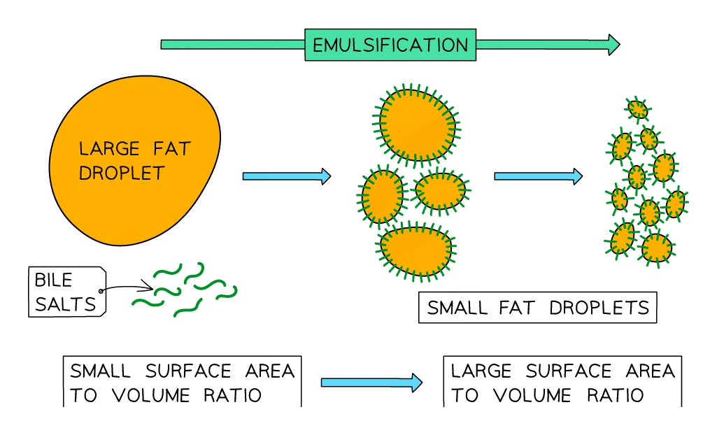
Lipase is a water-soluble enzyme, but lipids are oily and form large globules in the watery contents of the gut. Lipase can only attack a lipid at its surface, so a single big lipid globule is digested very slowly.
Bile salts in bile break each large lipid globule into many tiny droplets, called emulsification. The total surface area of all the small droplets is enormous compared to one big globule, so lipase has far more surface to attack, and lipid digestion speeds up dramatically.
Emulsification is mechanical digestion, not chemical digestion. The lipid molecules themselves are not broken; only the size of the droplets has changed.
9. Absorption in the small intestine
Once digestion is complete, the small soluble products (glucose, amino acids, fatty acids and glycerol) need to be absorbed into the blood. This happens in the ileum, the long second part of the small intestine.
Adaptations of the small intestine for absorption
The ileum is brilliantly adapted to take in the maximum amount of dissolved food in the shortest time:
- Very long (around 6 metres in an adult): the food spends a long time in contact with the absorbing surface
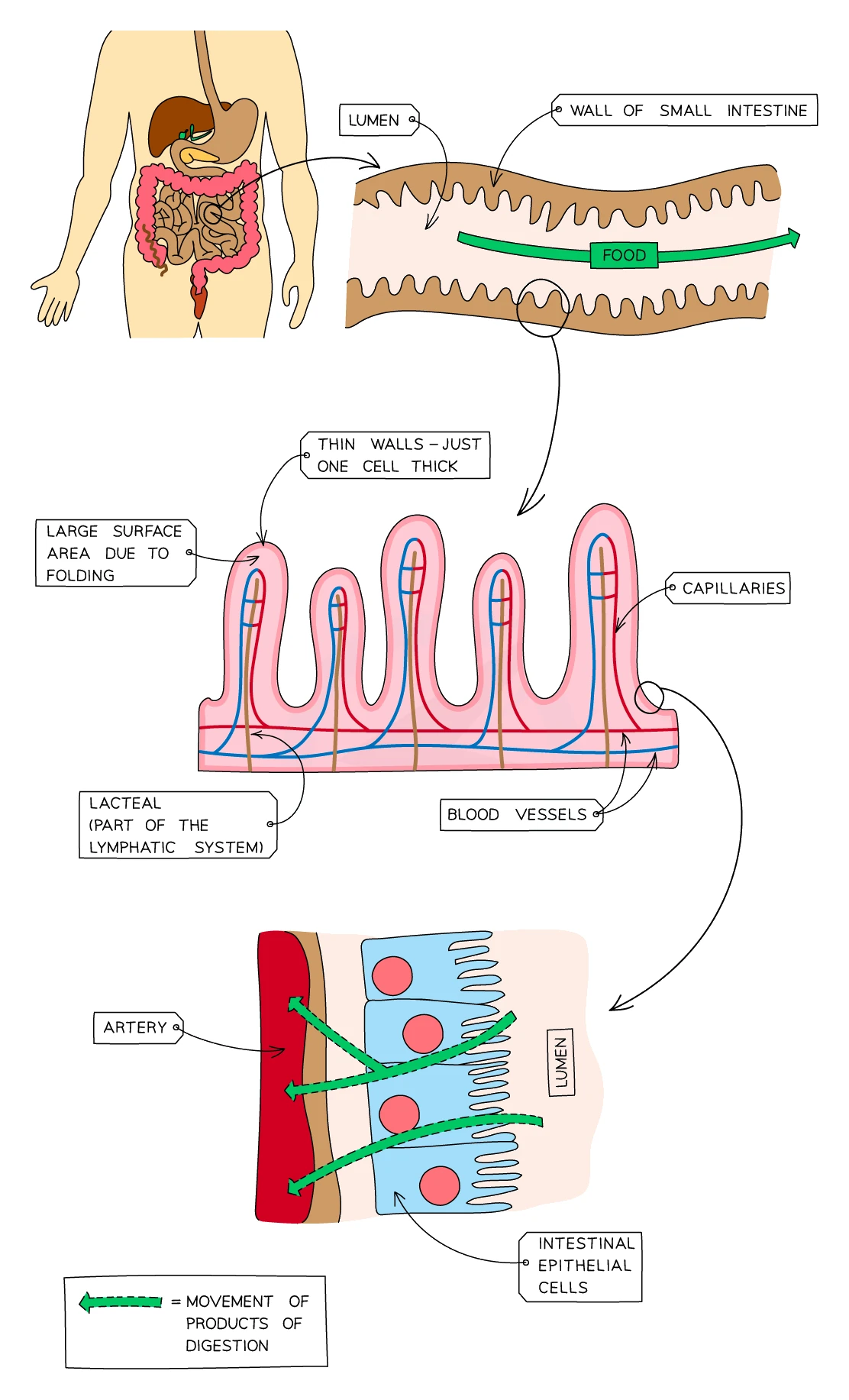
- Inner surface folded into millions of tiny finger-like projections called villi: each villus is about 1 mm long
- The cells covering each villus have their surfaces folded into thousands of even tinier
- Combined, the villi and microvilli increase the inner surface area to roughly 250 m², about the size of a tennis court
Structure of a villus
Each villus has:
- A thin wall just one cell thick (a single layer of epithelial cells), so the diffusion distance for absorbed molecules is very short
- A dense network of blood capillaries running just inside the wall, which carries away the glucose and amino acids as soon as they are absorbed. This keeps the concentration gradient between the gut and the villus steep, maintaining fast absorption
- A (a small lymph vessel) running up the centre, which absorbs the fatty acids and glycerol and carries them into the lymphatic system
Most digested food is absorbed by diffusion (down its concentration gradient into the blood). When the gradient runs out, active transport is used to absorb the last molecules against the gradient.
Exam tip
Pair every villus feature with its reason
When a question asks how the small intestine suits absorption (4 marks, set once in these papers), the scheme pays only for a feature joined to its purpose: the lining is folded into villi covered in microvilli, increasing the surface area; the wall is one cell thick, a short diffusion distance; capillaries absorb glucose and amino acids and keep the concentration gradient steep; the lacteal absorbs lipid. "Villi" or "thin wall" alone earns nothing. Multiple-choice items in two papers ask for the microvillus on a cell drawing, or the part of the gut with villi: the small intestine.
10. Saprotrophic nutrition in fungi
Fungi do their digestion outside their own body. This style of feeding is called saprotrophic nutrition:
- The fungus grows a tangled mesh of thread-like cells called hyphae (collectively the mycelium) into a piece of dead organic matter (a fallen leaf, an old log, a slice of bread).
- The hyphae secrete digestive onto the surrounding food. Carbohydrases, proteases and lipases are released, just like the ones in your gut.
- The enzymes break down the large food molecules into small soluble ones right there in the substrate.
- The fungus then absorbs the small soluble molecules through the cell wall of each hypha.
Because they break down dead material and recycle the nutrients, fungi are essential decomposers in every ecosystem. Without them, dead leaves, animals and other waste would simply pile up.
11. Core practical: measuring the energy content of food
Principle
You burn a known mass of food directly below a known volume of water. The heat from the burning food warms the water. By measuring the temperature rise, you can calculate the energy released, using the specific heat capacity of water (4.2 J for each gram of water raised by 1 °C).
Apparatus
- A boiling tube and clamp stand
- Measuring cylinder
- A thermometer (reading to at least 0.1 °C)
- Mass balance
- A mounted needle (to hold the food sample)
- Bunsen burner (to set the food alight)
- Small samples of food to test (popcorn, peanut, crisp, pasta, etc.)
Method
- Use the measuring cylinder to add 25 cm³ of cold water to the boiling tube. Clamp the boiling tube vertically.
- Record the starting temperature of the water.
- Find the mass of the food sample on the balance.
- Push the food sample onto the mounted needle.
- Set fire to the food with the Bunsen burner, then quickly hold the burning sample about 2 cm under the boiling tube. Keep it there until the food has finished burning.
- Stir the water gently with the thermometer and record the highest temperature the water reaches.
- Repeat for at least two more pieces of each food to get a mean.
Calculation
energy per gram of food (J/g) = (mass of water in g × temperature rise in °C × 4.2) ÷ mass of food in g
Where:
- The 4.2 is the specific heat capacity of water (4.2 J/g/°C)
- 1 cm³ of water has a mass of 1 g, so 25 cm³ of water = 25 g
Worked example
Energy per gram of a food from the temperature rise
A student burns a 0.45 g piece of pasta under a boiling tube holding 25 cm³ of water. The water warms from 21.0 °C to 39.0 °C. Calculate the energy released per gram of pasta.
Solution:
- Mass of water: 25 cm³ has a mass of 25 g
- Temperature rise: 39.0 − 21.0 = 18.0 °C
- Energy released = 25 × 18.0 × 4.2 = 1890 J
- Energy per gram = 1890 ÷ 0.45 = 4200 J/g (4.2 kJ/g)
The true value is higher, because some of the heat never reaches the water.
Limitations and how to improve
- Heat is lost to the air, not all of it reaches the water. The measured energy is always lower than the true energy in the food.
- Use a calorimeter (a copper container with insulation around the water) or wrap aluminium foil around the boiling tube to reduce heat loss.
- The food may not burn completely, especially if it goes out before all the energy is released.
- Relight the food repeatedly until it will no longer catch fire.
- Different foods catch fire at different rates, so the time held under the tube is hard to control.
- Use a fixed apparatus (such as a clamped sample holder) to standardise the distance and position.
Common exam question
Describing or improving the food-burning experiment
Question: Describe how to compare the energy content of foods by burning them, explain how the readings give the energy per gram, or say why the value is too low and how to improve the apparatus (2–5 marks).
Set in 4 of the 23 papers, all on Paper 2. A valid comparison needs the same mass of food, the same volume of water, the food relit under the tube until it will not burn, the temperature rise and repeats. Energy = temperature rise × 4.2 × mass of water, divided by the mass of food; writing it can earn most method marks. The value is too low because heat is lost to the air and in moving the food, combustion is incomplete and the water is not stirred. Explaining why a calorimeter gives a better value is marked in pairs, change plus reason: a lid or insulation to cut heat loss, a stirrer for even heating, and burning in oxygen for complete combustion. A two-mark list of improvements credits each change alone, including a fixed food position.
12. Immobilised industrial enzymes
Enzymes are also used in industry to speed up reactions outside the body. A common trick is to immobilise the enzyme so it can be reused.
What "immobilised" means
The enzyme is trapped inside small beads (typically beads of alginate, a jelly-like substance). The beads are then packed into a column. A liquid containing the substrate is poured through the column. The substrate diffuses into the beads, meets the enzyme, gets converted into product, and the product flows out of the bottom of the column.
The enzyme stays inside the beads and can be used again and again, instead of being washed away with the product.
Example: lactose-free milk
Many adults are lactose intolerant: they no longer make enough of the enzyme lactase, so the lactose (the sugar in milk) passes undigested into the gut and causes bloating, cramps and diarrhoea.
Lactose-free milk is made by running ordinary milk through a column packed with immobilised lactase. As the milk flows through, the trapped lactase splits the lactose into glucose and galactose, which are absorbed easily. The milk leaving the column is safe for lactose-intolerant people and tastes slightly sweeter, because glucose is sweeter than lactose. Because the enzyme stays in the beads, one column can treat milk continuously.