1. What homeostasis is
Homeostasis is the maintenance of a constant internal environment in the body, despite changes in the outside environment
Body cells only work properly within a narrow range of conditions. Enzymes have a fixed optimum temperature and pH; cell membranes only function inside a narrow range of solute concentrations. If conditions drift outside this safe range, the body's chemistry breaks down. A drop in body temperature of more than a few degrees can be fatal, as can a drop in blood glucose to zero or a 30% loss of body water.
Homeostasis is the collection of mechanisms that keep these conditions stable. The major variables that the body keeps constant include:
- Core body temperature (around 37 °C)
- Blood glucose concentration (around 4–7 mmol/L)
- Water content of the blood
- Salt (ion) concentration of the blood
- Blood pH (around pH 7.4)
- Levels of metabolic waste (urea, CO₂)
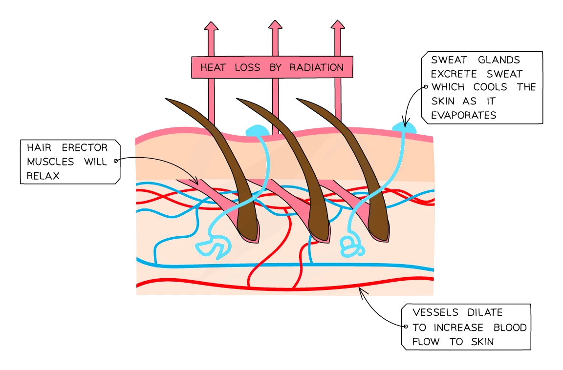
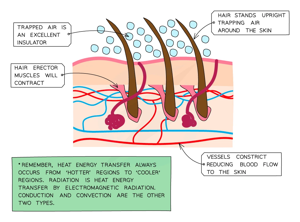
The mechanisms covered in detail in this topic are blood glucose regulation and osmoregulation (water balance). Temperature regulation by the skin was covered in topic 10 (Coordination and Response), and water reabsorption by the kidneys was covered in topic 9 (Excretion). All of them are examples of the same general principle: .
2. Negative feedback
The body keeps each variable stable by using a negative feedback loop. The idea is simple: any change away from the normal level triggers a response that reverses the change, bringing the variable back to normal.
A negative feedback loop has four components:
- A norm (the desired set point, e.g. 37 °C or 5 mmol/L glucose)
- A that detects when the variable has drifted away from the norm
- A that decides what corrective action to take
- An that carries out the correction
The crucial feature is that the correction is opposite to the change. If the variable goes up, the response brings it back down; if it goes down, the response brings it back up.
A simple analogy
A central-heating thermostat works the same way:
- The thermostat is set to (say) 20 °C.
- A sensor measures the room temperature.
- If the room cools below 20 °C, the thermostat switches the heater ON and the temperature rises.
- If the room warms above 20 °C, the thermostat switches the heater OFF and the room cools down.
The body works exactly like this for many of its variables. Most of the body's "thermostats" sit in the , a small region at the base of the brain that monitors the blood as it flows past.
The opposite: positive feedback
There is also , where a change triggers a response that amplifies the change. This is rare in the body and is usually unhelpful for maintaining stability. Childbirth (where stretching of the cervix triggers more contractions, which stretch the cervix further) is one of the few useful examples. For homeostasis, the body relies almost entirely on negative feedback.
3. Blood glucose regulation
Glucose is the main fuel for respiration, and every cell in the body needs a constant supply. The brain especially relies on it. The body keeps the blood glucose concentration within a narrow range: roughly 4 to 7 mmol per litre of blood.
Why blood glucose has to stay in range
- Too high a blood glucose pulls water out of cells by osmosis (because the blood is more concentrated than the inside of the cells). Cells become dehydrated and stop working. Sustained high blood glucose damages blood vessels, kidneys, nerves and eyes.
- Too low a blood glucose starves the cells of fuel. The brain is hit first, because it cannot store glucose. Symptoms range from confusion and dizziness to seizures, coma and death.
Two hormones, two opposite jobs
Two hormones from the pancreas work as a pair to keep blood glucose in range:
- Insulin is released when blood glucose is too high. Insulin lowers blood glucose.
- Glucagon is released when blood glucose is too low. Glucagon raises blood glucose.
The pancreas contains specialised endocrine cells that constantly monitor the glucose concentration of the blood flowing through it, releasing the appropriate hormone as conditions change.
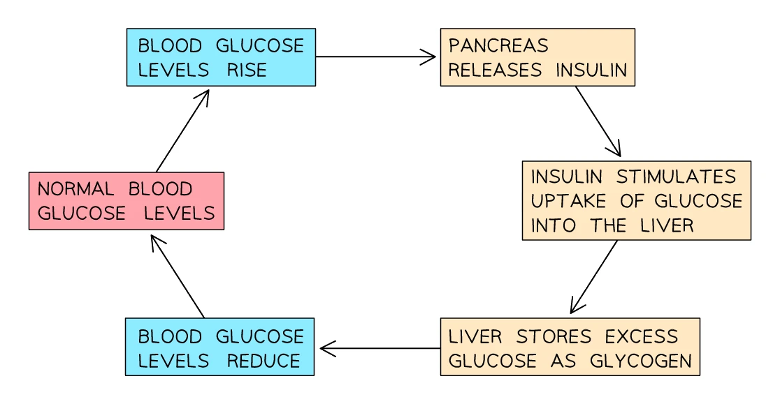
When blood glucose rises (after a meal)
After you eat a meal, especially one with lots of carbohydrates, glucose is absorbed from the small intestine and blood glucose climbs. The negative feedback loop kicks in:
- The pancreas detects the rise in blood glucose.
- The pancreas releases the hormone into the blood.
- Insulin travels to the liver and muscle cells, where it triggers two responses:
- Liver and muscle cells take up glucose from the blood
- The glucose is converted into , an insoluble storage molecule, and stored inside the cells
- Blood glucose falls back to normal.
- The pancreas stops releasing insulin.
When blood glucose falls (between meals, or during exercise)
Between meals (or during exercise, where the muscles are using glucose fast), blood glucose can drop too low. The opposite loop runs:
- The pancreas detects the fall in blood glucose.
- The pancreas releases the hormone into the blood.
- Glucagon travels to the liver, where it triggers the reverse of insulin's action:
- The liver breaks down its stored glycogen back into glucose
- The glucose is released into the blood
- Blood glucose rises back to normal.
- The pancreas stops releasing glucagon.
The pair of hormones acts like a thermostat: insulin to cool things off when glucose is high, glucagon to warm things up when glucose is low. Most of the time both hormones are at low levels and only one becomes active when needed.
| Situation | Pancreas releases | Target organs | What happens | Result |
|---|---|---|---|---|
| Blood glucose too high | Insulin | Liver and muscles | Glucose taken up and stored as glycogen | Blood glucose falls to normal |
| Blood glucose too low | Glucagon | Liver | Glycogen broken down back into glucose, released into blood | Blood glucose rises to normal |
Common exam question
Explaining how insulin lowers blood glucose
Question: Explain the role of insulin in the body, or complete a passage about what happens when the glucose concentration of the blood rises (2–4 marks).
Asked in 2 of the 23 papers, with one mark per link in the chain: a high blood glucose concentration makes the pancreas release insulin; insulin causes the liver and muscles to take up glucose and convert it to glycogen; so blood glucose falls. The two-mark explain question takes any two links; the passage version wants the four nouns (insulin, pancreas, liver or muscles, glycogen), one mark each.
Three words are policed. Glucose is converted into glycogen, never "broken down" into it, and the store is glycogen: "glucagon" for the storage substance is rejected. "Insulin controls blood glucose" is ignored, so say that it reduces it. The same facts fill a hormone table row (insulin, pancreas, converts blood glucose into glycogen) and a which-gland multiple-choice question in two more papers.
4. Diabetes
Diabetes mellitus is a chronic condition in which the body fails to control its blood glucose properly, so glucose levels rise to dangerously high values after a meal and stay high. There are two main forms: Type 1 and Type 2.
Type 1 diabetes
In Type 1 diabetes, the body's own immune system has attacked and destroyed the insulin-producing cells in the pancreas. The pancreas can no longer make insulin, so there is nothing to stop blood glucose climbing after a meal.
- Cause: an autoimmune attack on the pancreas. The reason this happens is not fully understood; genetics and viral infections in childhood are both thought to play a role.
- Usually appears in childhood or young adulthood.
- Not preventable and not linked to lifestyle.
- Treatment: lifelong injections before meals (insulin cannot be taken as a tablet because the stomach would digest it). The dose is adjusted based on what the patient eats and how active they are. Modern patients often use an insulin pump that delivers a continuous dose plus extra at meals, or a continuous glucose monitor that helps fine-tune the doses.
- Patients also have to:
- Monitor blood glucose regularly with a finger-prick test
- Eat a controlled carbohydrate diet with regular meals
- Exercise regularly (but with awareness, since exercise lowers blood glucose)
Exam tip
Diabetes questions test reasoning, not recall
Only 1 of the 23 papers has asked about diabetes itself (elsewhere it is a one-word answer in diet or heart questions), and the question defined the condition: the marks were for reasoning. Insulin is injected rather than swallowed because it is a protein, so it would be digested by protease in the stomach or small intestine (three points for two marks; "denatured in the stomach" is also credited). Exercise is monitored because it uses up glucose, so blood glucose could fall too low; the credited diet change is limiting carbohydrate or sugar (starch in place of sugar is accepted).
Type 2 diabetes
In Type 2 diabetes, the pancreas still makes insulin (at least to begin with), but the body cells stop responding to it properly. The cells become insulin-resistant: they no longer take up glucose efficiently in response to the hormone, so blood glucose stays high.
- Cause: strongly linked to lifestyle factors including obesity, lack of physical activity, and a diet high in sugar and processed carbohydrates. There is also a genetic predisposition in some families.
- Usually appears in adulthood, although increasingly in younger people because of rising rates of childhood obesity.
- Mostly preventable with lifestyle changes.
- Treatment:
- Lifestyle changes first: weight loss, more exercise, lower-carbohydrate diet, eating smaller more frequent meals
- Tablets (such as metformin) that help the body's cells respond to insulin better, or that slow down glucose absorption from the gut
- Insulin injections are sometimes needed eventually, if the pancreas wears out over many years
Side-by-side comparison
| Type 1 | Type 2 | |
|---|---|---|
| Cause | Autoimmune destruction of insulin-producing pancreatic cells | Body cells become resistant to insulin; pancreas eventually wears out |
| Insulin production? | No, or very little | Yes (at least at first) |
| Age of onset | Usually childhood / young adult | Usually adult (over 40), but rising in younger people |
| Linked to lifestyle? | No | Yes: obesity, inactivity, poor diet |
| Preventable? | No | Largely yes |
| First-line treatment | Insulin injections | Lifestyle changes, then tablets |
| Approximate share of cases | ≈ 10% of diabetics | ≈ 90% of diabetics |
Symptoms and complications
Symptoms of untreated or poorly-controlled diabetes are similar for both types and reflect the high blood glucose:
- Frequent urination: high blood glucose forces the kidneys to excrete some of the excess in urine. To carry the glucose out, the kidneys also lose extra water, so urine volume rises.
- Constant thirst: the body needs to replace all the water lost in urine.
- Tiredness: glucose cannot enter the cells properly, so cells are short on fuel.
- Sugar in the urine: a positive Benedict's test on a sample of urine is a classic warning sign.
- Slow wound healing and frequent infections.
- Unexplained weight loss (especially in Type 1, where the body starts breaking down fat and muscle for energy).
Long-term complications of uncontrolled diabetes include damage to the eyes (leading to blindness), kidneys (leading to kidney failure), nerves (leading to loss of sensation, especially in the feet), and blood vessels (leading to heart attacks, strokes, and poor circulation in the legs).
5. Osmoregulation: controlling water balance
Osmoregulation is the control of the water content and solute concentration of the body fluids, keeping them within a narrow range
Body cells only work properly when surrounded by tissue fluid with the right water concentration. If the surrounding fluid is too concentrated (too little water), water leaves cells by osmosis and they shrivel. If the surrounding fluid is too dilute (too much water), water enters cells by osmosis and they swell or burst.
The body's water balance therefore has to be tightly controlled in spite of the constant inputs and outputs of water:
| Water IN | Water OUT |
|---|---|
| Drinks | Urine |
| Food | Sweat (skin) |
| Aerobic respiration (a small amount) | Breath (lungs) |
| Faeces (a small amount) |
Most water inputs and outputs are roughly self-balancing: if you drink more, you produce more urine. But to keep blood water concentration steady from minute to minute, the body adjusts how much water the kidneys keep back from the urine.
The role of ADH
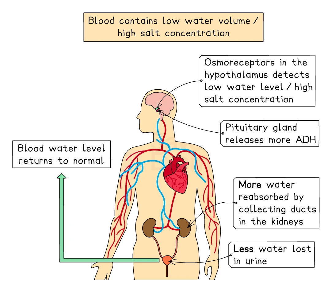
The hormone ADH (antidiuretic hormone), released by the pituitary gland in the brain, controls the kidney's water reabsorption. ADH was covered in detail in topic 9 (Excretion); the summary here is enough for the homeostasis perspective.
The negative feedback loop:
When blood water is too LOW (dehydrated, blood too concentrated):
- Osmoreceptors in the detect the increase in blood solute concentration.
- The hypothalamus signals the pituitary gland to release more ADH.
- ADH makes the kidney's collecting ducts more permeable to water.
- More water is reabsorbed from the filtrate back into the blood.
- A small volume of concentrated (dark) urine is produced.
- Blood water concentration rises back to normal.
When blood water is too HIGH (over-hydrated, blood too dilute):
- Osmoreceptors detect the decrease in blood solute concentration.
- The pituitary releases less ADH.
- The collecting ducts become less permeable to water.
- Less water is reabsorbed; more stays in the filtrate.
- A large volume of dilute (pale) urine is produced.
- Blood water concentration falls back to normal.
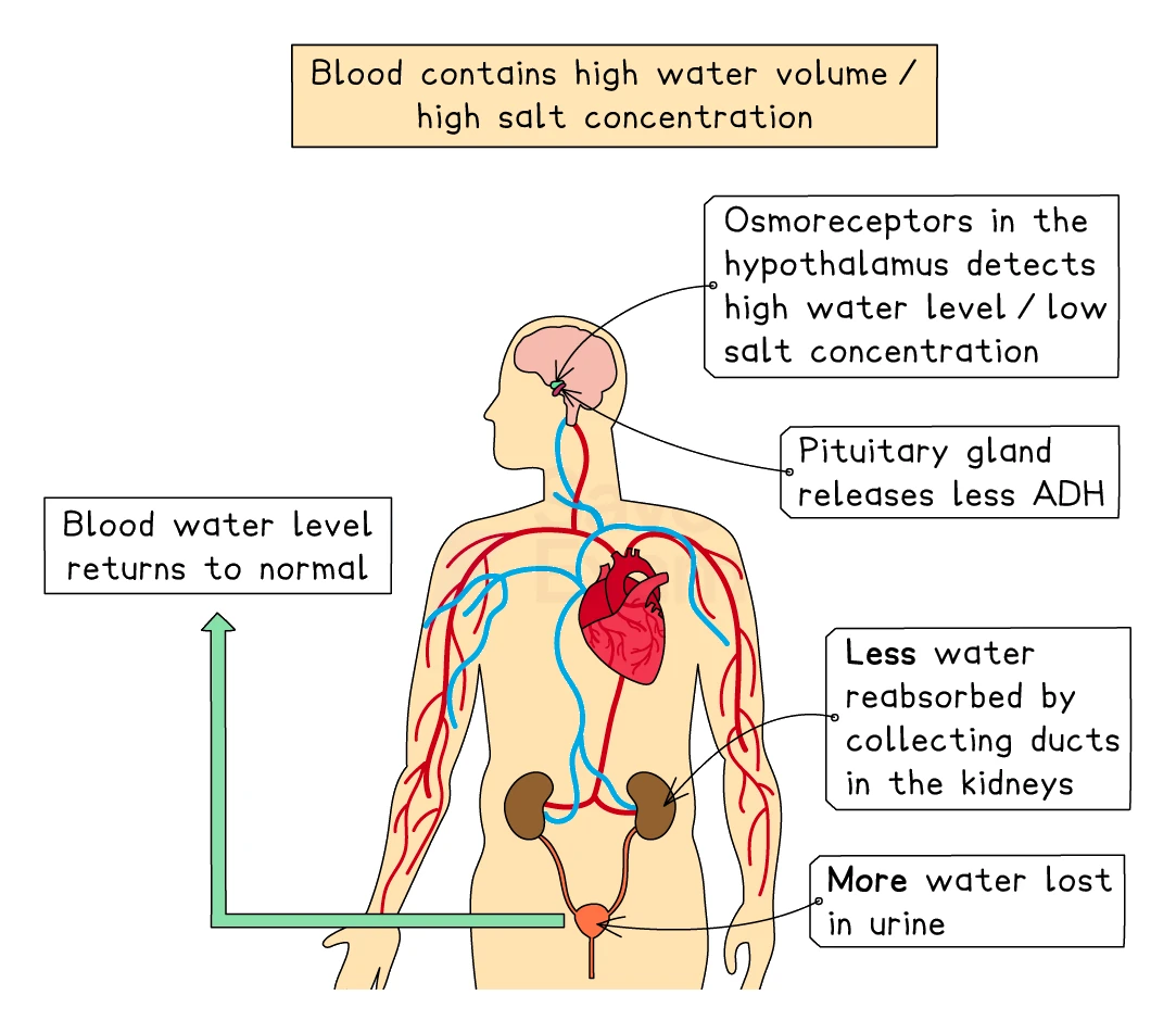
You will also feel thirsty when blood water is low, prompting you to drink more water. The thirst response and the ADH response work together.
Why drinking very salty water makes you more thirsty
Sea water is much more concentrated than blood plasma. If you drink sea water, the salt enters your blood and raises the blood's solute concentration even further. The body responds as if dehydrated: it tries to produce concentrated urine to get rid of the extra salt, but to do so it has to use up even more of the body's water. The net effect is to make the body more dehydrated than before. This is why drinking sea water when stranded does not save you and can speed up your death from thirst.
6. Bringing it together
The three homeostatic systems covered here (or referenced from other topics) all use the same general structure:
| Variable | Receptor | Coordinator | Effector | Hormone(s) involved |
|---|---|---|---|---|
| Body temperature | Temperature receptors in skin and hypothalamus | Hypothalamus | Skin (sweat glands, arterioles, hair muscles), skeletal muscle (shivering) | None directly; nervous coordination |
| Blood glucose | Cells in the pancreas | Pancreas | Liver, muscles | Insulin (lowers), glucagon (raises) |
| Blood water content | Osmoreceptors in hypothalamus | Hypothalamus → pituitary gland | Kidneys (collecting ducts) | ADH |
In each case the principle is the same: detect the change, respond with the opposite, restore the norm. Once you understand this pattern, every homeostatic system in the body fits the same template.