1. Key genetic terms
Inheritance is a topic where the right words matter. Most exam questions on genetics rely on you knowing what a gene, an allele, a genotype and a phenotype are, and being able to use these terms precisely.
The big eight
| Term | What it means |
|---|---|
| DNA | The chemical that carries all the genetic information of an organism. Stands for deoxyribonucleic acid |
| Chromosome | A long, tightly-coiled molecule of DNA found in the nucleus. Humans have 23 pairs in every body cell |
| Gene | A short section of DNA on a chromosome. Each gene codes for one specific protein |
| Allele | One of the different versions of the same gene. A gene might have two or more alleles |
| Genotype | The combination of alleles an individual has for a particular gene (e.g. BB, Bb, bb) |
| Phenotype | The observable characteristic that results from a particular genotype (e.g. brown eyes, blood group A) |
| Dominant | An allele whose effect is seen in the phenotype even when only one copy is present. Written as a capital letter (e.g. B) |
| Recessive | An allele whose effect is only seen in the phenotype when both copies are present. Written as a lowercase letter (e.g. b) |
Common exam question
Defining a key genetic term
Question: State what is meant by the term gene, allele, genome, recessive allele, codominant or mutation (1 mark).
Asked in 10 of the 23 papers, usually as the first part of a longer genetics question. Each definition is one mark. A gene is a length of DNA coding for one protein. An allele is a different form or version of a gene. The genome is all the DNA, or all the genes, of an organism; when a question asks how a gene differs from a genome, both definitions are needed for the single mark. A recessive allele is one that is only expressed when two copies are present (in a homozygote), or when no dominant allele is present: either wording is credited. Codominant alleles are both expressed in the phenotype of a heterozygote, which shows a third phenotype (blood group AB, or roan cattle). A mutation is a rare, random change to the DNA or to a gene.
Homozygous and heterozygous
The two alleles you carry for a given gene can be the same or different:
- Homozygous: both alleles are the same (e.g. BB or bb)
- Heterozygous: the two alleles are different (e.g. Bb)
If you are heterozygous and one allele is dominant, the dominant version is the one you show in your phenotype. The recessive allele is still there in your genotype, but it is masked by the dominant one.
An example: the PTC tasting gene. Some people can taste a bitter chemical called phenylthiocarbamide (PTC, found naturally in some vegetables like sprouts); others can't taste it at all. The ability to taste PTC is controlled by one gene with two alleles:
- T = can taste PTC (dominant)
- t = cannot taste PTC (recessive)
| Genotype | Possible alleles | Phenotype |
|---|---|---|
| TT | T from each parent | Taster (homozygous dominant) |
| Tt | T from one parent, t from the other | Taster (heterozygous; T masks t) |
| tt | t from each parent | Non-taster (homozygous recessive) |
Note that two different genotypes (TT and Tt) produce the same phenotype (taster). The only way to be a non-taster is to have two copies of the recessive allele.
Common exam question
Genotypes behind a dominant phenotype
Question: Give the possible genotypes of an individual with the dominant characteristic, or explain how a cross would show which genotype it has (1–2 marks).
Set in 3 of the 23 papers. For the possible genotypes give both: homozygous dominant and heterozygous (TT and Tt). Adding the homozygous recessive genotype to that list loses the mark. To tell the two apart, breeders use a test cross with a homozygous recessive partner (tt): if every offspring shows the dominant characteristic the parent was homozygous, while any recessive offspring proves it was heterozygous, because a recessive offspring needs a recessive allele from both parents. The two marks come from any two of those three points: the cross with the homozygous recessive, and what each outcome shows.
Other terms you should know
- Genome: the whole of an organism's DNA, taken together. Every chromosome, every gene, every base. Sequencing the entire human genome was completed in 2003.
- Diploid: a cell with two complete sets of chromosomes (e.g. a human body cell with 23 pairs = 46 chromosomes). Written as 2n.
- Haploid: a cell with one complete set of chromosomes (e.g. a human sperm or egg with 23 chromosomes). Written as n.
- Variation: differences between individuals of the same species. Variation can be genetic (caused by different alleles), environmental (caused by lifestyle, diet, accidents), or a mix of both.
2. DNA structure
DNA (deoxyribonucleic acid) is the molecule that stores all the genetic information in every living cell. Its structure was worked out in 1953 by Watson, Crick, Franklin and Wilkins.
The double helix
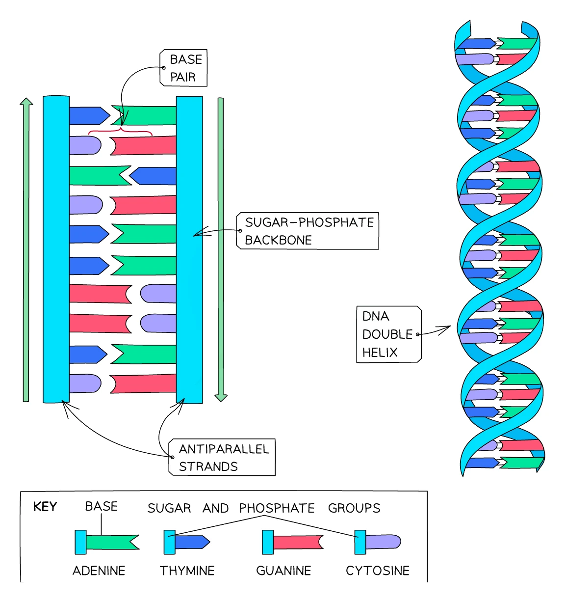
DNA is built as a double helix: two long strands wound around each other in a regular spiral, like a twisted ladder. Each strand is made of a chain of smaller units called nucleotides.
A nucleotide has three parts:
- A phosphate group (forms part of the strand backbone)
- A deoxyribose sugar (also part of the backbone)
- A nitrogenous base (the variable part)
The phosphates and sugars alternate along the outside of each strand, forming the sugar-phosphate backbone. The bases stick inwards from the strands and form the rungs of the ladder.
The four bases and how they pair
DNA has only four different bases:
- A (adenine)
- T (thymine)
- G (guanine)
- C (cytosine)
On the two strands of the helix, the bases pair up across the middle of the ladder by following strict complementary base-pairing rules:
- A always pairs with T
- G always pairs with C
This means that if you know the order of bases on one strand, you can work out the order on the other. For example, if one strand reads A-T-G-C-A, the partner strand must read T-A-C-G-T.
The two strands are held together by hydrogen bonds between the paired bases. These are weak enough to be broken easily (so the strands can separate during DNA replication and protein synthesis) but strong enough in total to keep the helix stable.
Genes are sections of DNA
A gene is just a length of DNA that codes for one specific protein. A typical human chromosome contains thousands of genes, each separated from the others by non-coding regions.
The sequence of bases in a gene determines the sequence of amino acids in the protein it codes for. The genetic code works in groups of three bases: every group of three bases (a triplet) codes for one specific amino acid.
3. RNA structure
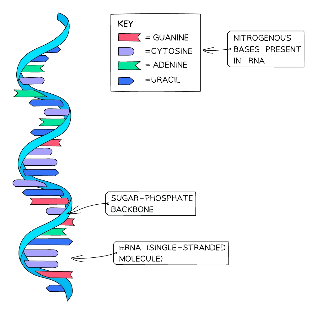
RNA (ribonucleic acid) is the messenger molecule that carries the instructions in a gene from the nucleus to the place where proteins are made. RNA is similar to DNA but with three important differences:
| Feature | DNA | RNA |
|---|---|---|
| Number of strands | Two (double helix) | One (single strand) |
| Sugar | Deoxyribose | Ribose |
| Bases used | A, T, G, C | A, U, G, C (uracil replaces thymine) |
| Length | Long (entire chromosomes) | Short (a copy of one gene) |
| Location | In the nucleus | Made in the nucleus, used in the cytoplasm |
In RNA, uracil (U) plays the role that thymine (T) plays in DNA, so the new pairing rules are:
- A pairs with U (in RNA-DNA pairing)
- G pairs with C
Common exam question
Writing a complementary DNA or mRNA sequence
Question: Give, or choose from four options, the base sequence of the other DNA strand, or of the mRNA made from a strand of DNA (1–2 marks).
Set in 4 of the 23 papers, always on Paper 2. Work base by base with the pairing rules: A with T and C with G across the two DNA strands, but A with U when the product is mRNA. When you write the sequence out for two marks, one wrong letter drops you to one mark, and so does writing T in an mRNA strand instead of U. The multiple-choice versions build their wrong options from exactly these slips (a DNA strand containing U, an RNA strand containing T, or a sequence that is simply not complementary), so check which molecule is asked for before you check the pairing. Three bases code for one amino acid, so a strand of nine bases codes for at most three.
There are several different types of RNA. The two most important for protein synthesis are:
- Messenger RNA (mRNA): a copy of a single gene that carries the code from the nucleus to a ribosome
- Transfer RNA (tRNA): brings the right amino acids to the ribosome in the right order
4. Protein synthesis: from gene to protein
The process of using a gene to build a protein has two main stages:
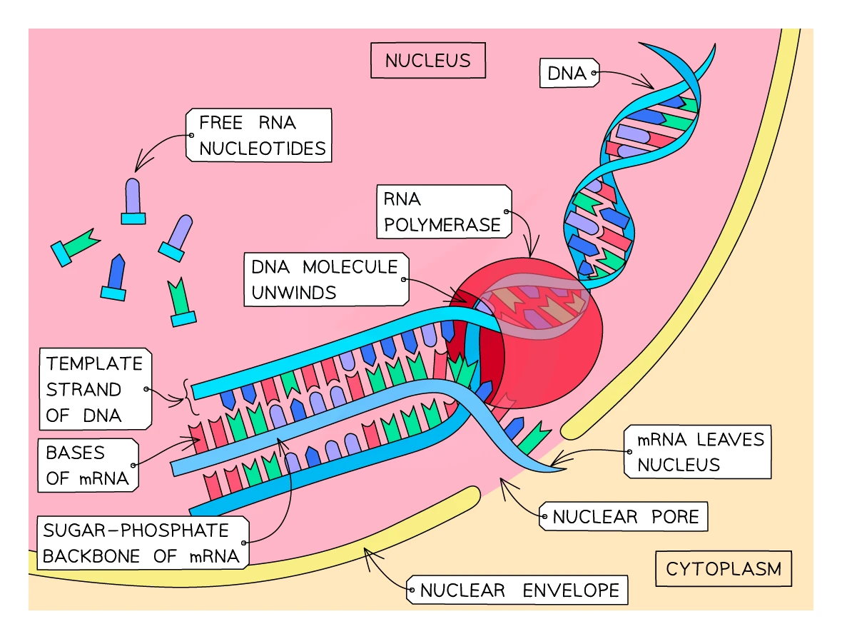
- Transcription: the DNA code of the gene is copied into an mRNA molecule, inside the nucleus.
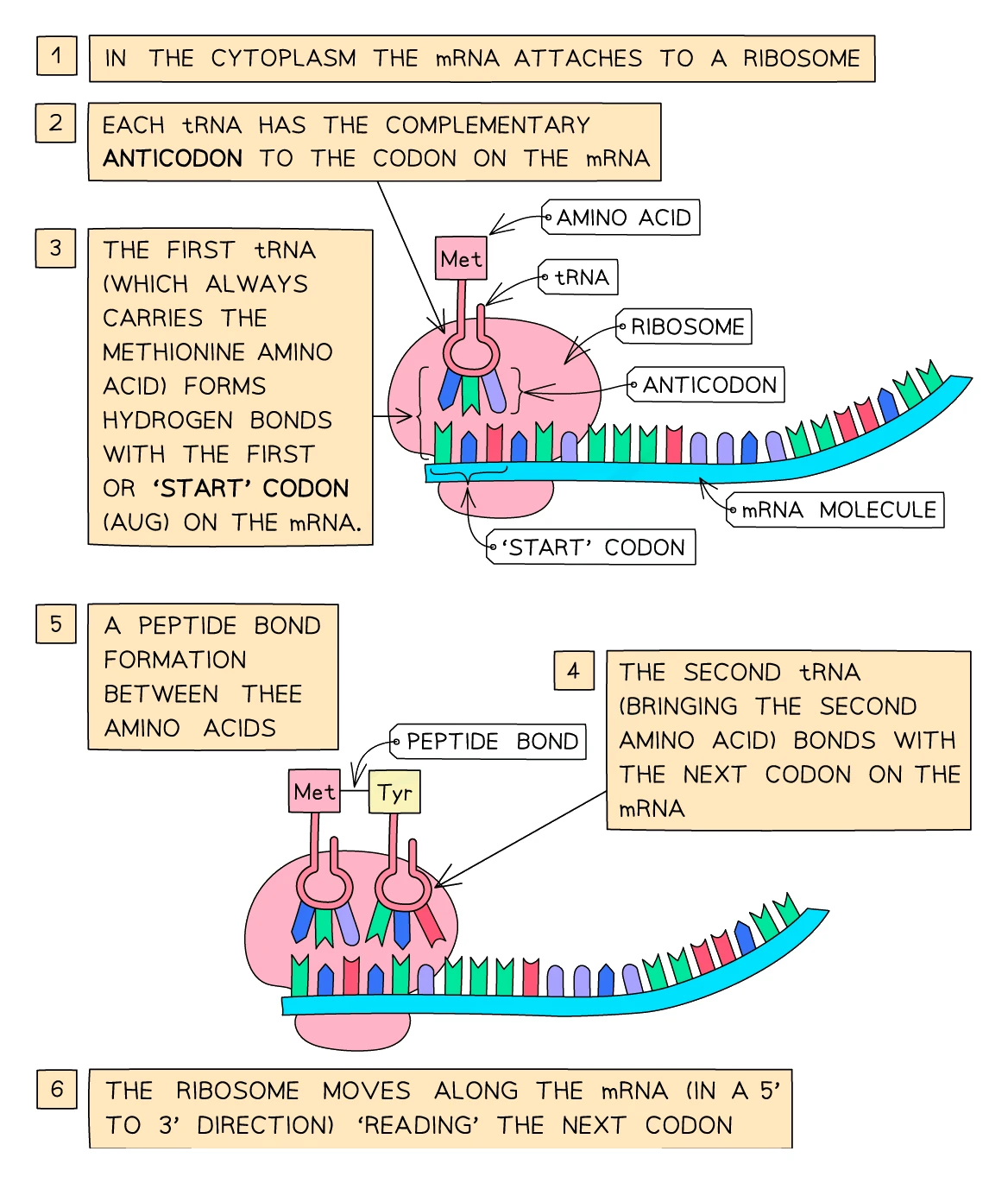
- Translation: the mRNA molecule is read by a ribosome, which uses the code to build the corresponding chain of amino acids in the cytoplasm.
Transcription (in the nucleus)
- The relevant section of DNA unwinds as hydrogen bonds between the complementary bases break.
- One strand of the gene acts as the template strand.
- Free RNA nucleotides in the nucleus line up against the template strand according to the complementary base-pairing rules (A pairs with U, T pairs with A, G pairs with C, C pairs with G).
- The RNA nucleotides are joined together into a single-stranded mRNA molecule.
- The mRNA detaches from the DNA, which rewinds back into a double helix.
- The mRNA molecule leaves the nucleus through a pore in the nuclear envelope.
So transcription produces an mRNA copy of the gene that can travel out of the nucleus to the protein-building machinery.
Translation (in the cytoplasm)
- The mRNA molecule arrives at a ribosome in the cytoplasm.
- The ribosome reads the mRNA three bases at a time. Each group of three bases is a and codes for one specific amino acid.
- In the cytoplasm, many free tRNA molecules are floating around. Each tRNA carries a specific amino acid at one end and has a triplet of bases () at the other end.
- A tRNA whose anticodon matches the first codon on the mRNA binds to it. Then a second tRNA binds to the next codon. The two amino acids carried by these tRNAs are joined together by a peptide bond.
- The ribosome moves along the mRNA one codon at a time. Each time, a new tRNA brings the matching amino acid, which is added to the growing chain.
- When the ribosome reaches a stop codon, translation stops. The completed chain of amino acids is released.
- The chain folds up into the specific 3D shape of the finished protein, which can now do its job (e.g. as an enzyme, antibody or structural protein).
The big picture
Gene (DNA) → mRNA (transcription, in the nucleus) → Protein (translation, on the ribosome)
The sequence of bases in DNA → determines the sequence of bases in mRNA → determines the sequence of amino acids in the protein → determines the shape and function of the protein → determines the characteristic of the organism.
Common exam question
Describing protein synthesis
Question: Describe the process of transcription, the stages of protein synthesis, the roles of RNA, or the differences between transcription and translation (4 marks).
Set as a 4-mark describe in 5 of the 23 papers, all on Paper 2, plus a 1-mark multiple-choice version in two more. The four marks come from a list of credited points, so write separate steps. Transcription: the DNA unwinds, mRNA is built as a copy of the DNA base sequence in the nucleus, and the mRNA leaves for a ribosome. Translation: the mRNA binds to the ribosome, tRNA brings amino acids, each anticodon pairs with a codon, and the amino acids are joined into a polypeptide. For differences, give each as a pair: nucleus against ribosome, DNA against mRNA as the starting molecule, mRNA against polypeptide as the product, no tRNA against tRNA. Two schemes ignore a bare "protein is made" for the last point: say amino acids are joined into a chain.
5. Monohybrid inheritance and Punnett squares
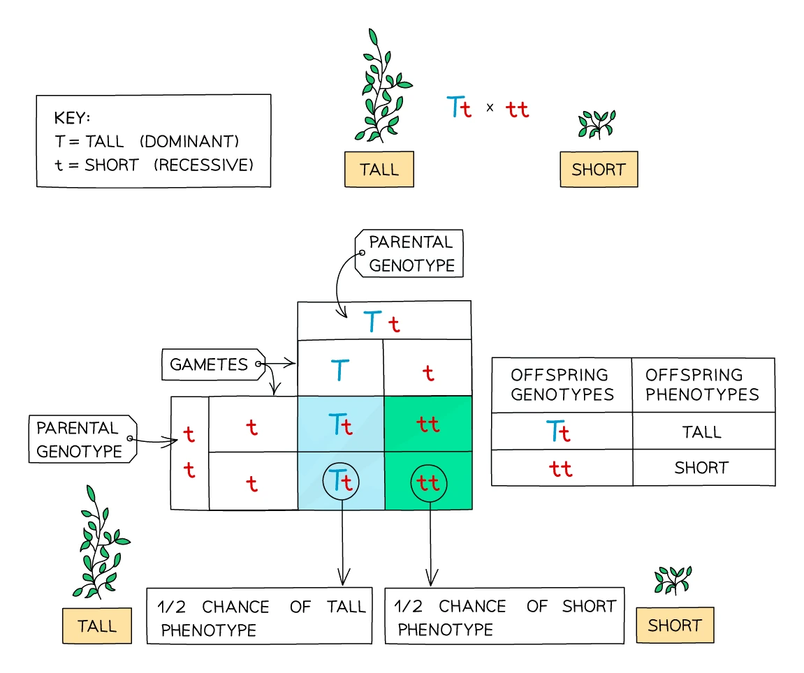
A monohybrid cross is a cross between two individuals looking at one gene with two alleles. The standard tool for predicting the outcome of a monohybrid cross is a .
How to set up a Punnett square
- Identify the parent genotypes. Use a clear letter for the gene, with the capital for the dominant allele and the lowercase for the recessive (e.g. T and t, or B and b; avoid letters like C/c and S/s where the case is hard to tell apart in handwriting).
- Split each parent's into the two gametes they can produce.
- Draw a 2×2 grid. Put one parent's possible gametes along the top, the other parent's possible gametes down the side.
- Fill in the four boxes of the grid by combining the alleles at the top and side of each box. These are the possible offspring genotypes.
- Work out the phenotype for each genotype using the dominant/recessive rules.
- State the ratio of phenotypes.
Common exam question
Drawing a genetic diagram for a monohybrid cross
Question: Draw a genetic diagram for the cross described, showing both parents' genotypes, their gametes, and the offspring genotypes and phenotypes (3–4 marks).
Asked in 11 of the 23 papers. Read the stem first: if parents with different phenotypes produce offspring that all look alike, the offspring show the dominant characteristic and are heterozygous (asked once for two marks: name it, then say all the offspring show it). Use one letter as capital and lower case (Rr): every scheme accepts it, some refuse two different letters, and none accepts X and Y. Most schemes give a mark each for the parental genotypes, gametes and offspring genotypes; four-mark versions add one for the offspring phenotypes (stated, or as a ratio) or for the probability asked for. A Punnett square earns them too; when phenotypes are asked for, label each genotype with its phenotype. With wrong parents, error carried forward usually earns one or two later marks, so finish the diagram.
From ratio to probability
Each new offspring is an independent event. The ratio from a Punnett square tells you the probability of each outcome for any single offspring.
For example, with Tt × Tt:
- Probability of a tall offspring = 3/4 = 0.75 = 75%
- Probability of a short offspring = 1/4 = 0.25 = 25%
If the parents have four offspring, the ratio will not always come out exactly 3:1. You might get 4 tall and 0 short, or 2 tall and 2 short. The 3:1 ratio only emerges accurately over many offspring. This is why pea plants are useful for these experiments, because a single plant can produce hundreds of seeds.
Common exam question
Comparing an observed ratio with the expected 3 : 1
Question: Calculate the offspring ratio in the form n:1, then explain why it differs from the expected ratio, or comment on results against the expected results (2–4 marks).
Set in 2 of the 23 papers. For the ratio, subtract to find the second group, write the unsimplified ratio (one mark on its own), then divide both sides by the smaller number to reach n:1, to one or two decimal places. For the explanation, state the expected ratio first (3:1 from two heterozygous parents, 1:1 for male to female), then that fertilisation is random, so which gametes fuse is down to chance (one mark each; "random fertilisation" alone earns both); a further mark comes from fewer of one type surviving or germinating. When comparing results, turn the counts into a ratio or percentage and set them beside the expected values: raw numbers alone earn nothing, while "close to 3:1, slightly fewer of one type than expected" earns the comparison marks.
Worked example
Genetic diagram for two heterozygous parents
Two plants are both heterozygous for stem length. The allele for long stem (W) is dominant over the allele for short stem (w). Both parents have genotype Ww. Predict the genotypes and phenotypes of their offspring.
Solution:
- Parental genotypes: Ww × Ww
- Parental gametes: W or w (from each parent)
- Punnett square:
| W | w | |
|---|---|---|
| W | WW | Ww |
| w | Ww | ww |
- Offspring genotypes: 1 WW : 2 Ww : 1 ww
- Offspring phenotypes: 3 long stem (WW and Ww) : 1 short stem (ww)
- Phenotype ratio = 3:1
6. Codominance: when both alleles show
In most monohybrid crosses, one allele is dominant and the other is recessive. But sometimes both in a heterozygote show their effects in the phenotype. This is called .
The ABO blood group system
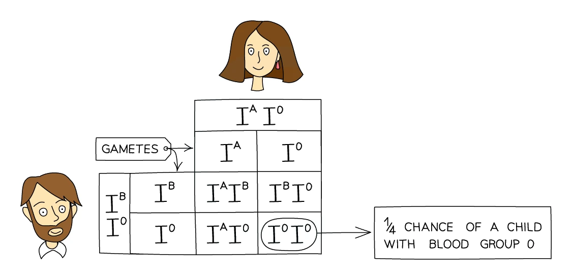
The classic example is the inheritance of human blood groups. The ABO gene has three alleles (not two):
| Allele | What it codes for |
|---|---|
| I^A | Production of antigen A on red blood cells |
| I^B | Production of antigen B on red blood cells |
| I^O | No antigen produced (no A and no B) |
The rules of dominance are:
- I^A and I^B are codominant with each other. If you have one of each, both antigens appear on your red blood cells.
- Both I^A and I^B are over I^O.
This gives four possible phenotypes (blood groups A, B, AB, O) from six possible genotypes:
| Genotype | Phenotype (blood group) |
|---|---|
| I^A I^A | A |
| I^A I^O | A |
| I^B I^B | B |
| I^B I^O | B |
| I^A I^B | AB (codominance: both A and B antigens) |
| I^O I^O | O |
7. Sex chromosomes and sex determination
The 23rd pair of human chromosomes is the sex chromosomes. The other 22 pairs are called autosomes and are the same in males and females.
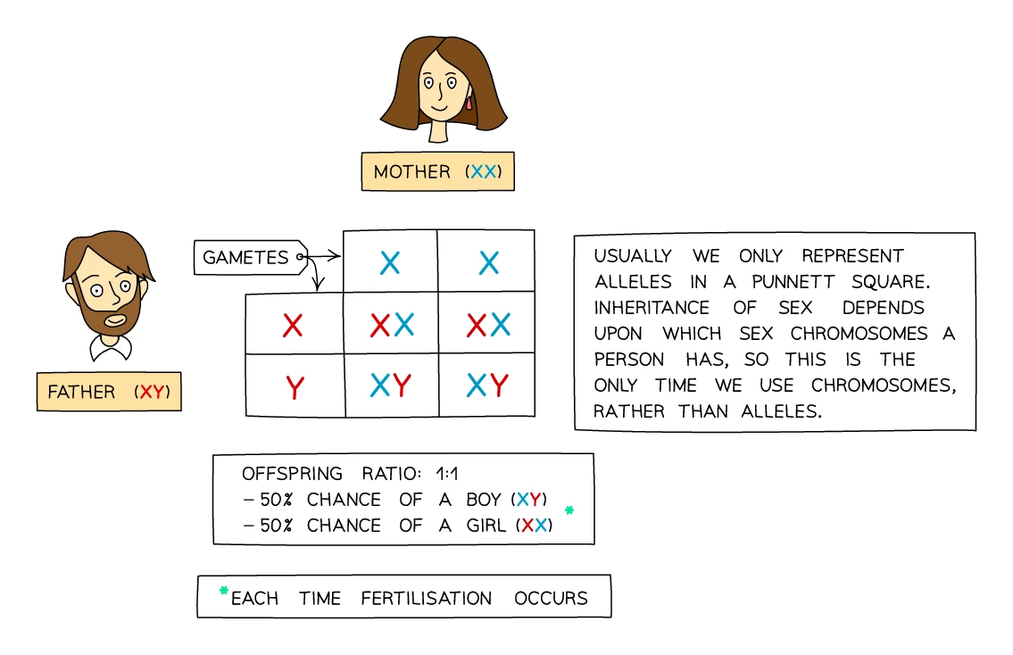
- Females have two X chromosomes (XX)
- Males have one X and one Y chromosome (XY)
The X chromosome is much larger than the Y and carries many more genes. The Y chromosome is short and carries relatively few genes, but it does contain the SRY gene that triggers the development of male reproductive organs in an embryo.
How sex is inherited
When gametes are made:
- A mother's eggs all carry an X chromosome (she has no Y to pass on).
- A father's sperm carry either an X or a Y, in a 50:50 mix.
So the father determines the sex of the child:
- If an X-carrying sperm fertilises the egg → XX → female
- If a Y-carrying sperm fertilises the egg → XY → male
Punnett square for inheritance of sex:
| X (from mother) | X (from mother) | |
|---|---|---|
| X (from father) | XX (female) | XX (female) |
| Y (from father) | XY (male) | XY (male) |
The ratio is 1 male : 1 female, so each new child has a 50% chance of being male and a 50% chance of being female.
Common exam question
Probability of a male offspring with a given phenotype
Question: Calculate the probability that an offspring of the cross is male and shows a stated characteristic (1–2 marks).
Asked in 6 of the 23 papers, always attached to the genetic diagram. Multiply two independent probabilities: 1/2 for male (or female), from the sex chromosomes, and the phenotype's share from your Punnett square (3/4 dominant and 1/4 recessive from two heterozygous parents; 1/2 each when a heterozygote is crossed with a homozygous recessive). So a male with the dominant phenotype is 1/2 × 3/4 = 3/8, accepted as 0.375 or 37.5%, and a male with the recessive phenotype is 1/8. For two marks the correct answer alone scores both, and 0.5 or 0.75 on its own scores one. When the probability is the final mark of the diagram question, most schemes award it only from a correct cross, so a wrong parental genotype usually costs it even though the gametes and offspring marks can survive by error carried forward.
8. Family pedigrees
A family pedigree is a diagram that traces how a particular characteristic (often a genetic disorder) passes through several generations of a family. Pedigree diagrams are useful for working out:
- Whether the characteristic is or
- The probability that a particular family member is affected, or could pass the condition on to their children
Pedigree symbols
Pedigree diagrams use standard symbols:
- Squares represent males; circles represent females
- Filled-in (shaded) shapes represent affected individuals (those showing the characteristic)
- Empty shapes represent unaffected individuals
- A horizontal line between a male and a female means they are a couple who have produced children
- A vertical line going down from a couple connects them to their children, who are shown side by side
Reading a pedigree
To work out whether a trait is dominant or recessive:
- If two unaffected parents have affected children, the trait must be recessive (because both parents are carriers but don't show the trait).
- If every affected child has at least one affected parent, the trait is likely to be dominant.
Common exam question
Reading genotypes from a family pedigree
Question: Use the pedigree to give the genotypes of named individuals, say how many are heterozygous, or draw a genetic diagram for a couple on it to find the probability of an affected child (1–4 marks).
Asked in 4 of the 23 papers, each time for a recessive condition. Work outwards from the affected individuals. Anyone affected is homozygous recessive. Both unaffected parents of an affected child must be heterozygous (carriers), and so must every unaffected child of an affected parent, because that child inherited one recessive allele. Counting the carriers this way answers the multiple-choice "how many are heterozygous" version. For the genetic diagram the couple is either two carriers, giving a 1/4 chance of an affected child, or a carrier with an affected partner, giving 1/2; it is marked like any other cross.
9. Mutations
A mutation is a rare, random change in the sequence of bases in a DNA molecule. Mutations happen continuously in every living cell, mostly without consequence.
Three types of mutation
There are three main ways the DNA sequence can change:
| Type | What happens | Effect |
|---|---|---|
| Substitution | One base is swapped for a different base | Only the codon containing the changed base is affected. The amino acid coded for by that codon may change, but the rest of the sequence is unaffected |
| Insertion | A new base is added into the sequence | Shifts every codon downstream of the insertion. Usually causes a much bigger change in the protein |
| Deletion | A base is removed from the sequence | Like insertion, shifts every codon downstream. Usually a major effect |
Most mutations don't matter
The vast majority of mutations have no effect on the organism. They can be neutral for several reasons:
- The mutation happens in a non-coding region of DNA (the bit between genes).
- The new codon still codes for the same amino acid (the genetic code has redundancy: many amino acids are coded for by several different codons).
- The new amino acid is so similar to the original that the protein's shape and function are unaffected.
Common exam question
Why a mutation may or may not change the phenotype
Question: Explain how a gene mutation can change the phenotype, or why most mutations have no effect on the phenotype or on an enzyme (3–5 marks).
Set in 5 of the 23 papers, all on Paper 2. For an effect, build the chain: a random change in the DNA base sequence, so the mRNA carries different codons, a different tRNA brings a different amino acid, the amino acid sequence changes, the protein folds into a different shape, and an enzyme's active site no longer fits its substrate. For no effect, give three separate reasons: several codons code for the same amino acid (as the codon table shows); a changed amino acid may leave shape and active site unaltered; the change may lie in non-coding DNA; or the mutated allele may be recessive and not expressed. Asked what increases the mutation rate, name ionising radiation, UV light or tar; tobacco and smoking also score, but infrared radiation does not.
When mutations do matter
Occasionally a mutation produces a real change in the protein, with consequences:
- Harmful produce a protein that does not work properly. Many genetic diseases are caused by mutations of this kind. Examples:
- Sickle cell anaemia: a single base substitution in the gene for haemoglobin changes one amino acid. The mutant haemoglobin sticks together in the wrong shape, deforming red blood cells into sickle shapes that block small blood vessels.
- Cystic fibrosis: a mutation in a gene called CFTR produces a faulty membrane protein. This causes thick sticky mucus to build up in the lungs and digestive system.
- Huntington's disease: an extra repetition of three bases in a gene called HTT produces a protein that damages brain cells in middle age.
- Beneficial mutations produce a protein that works better, or gives a new function. If the new variant helps the organism survive or reproduce, it can spread through the population by natural selection (covered in topic 14).
- Cancer is caused by mutations in the genes that normally control cell division. The mutated cells divide uncontrollably, forming a tumour.
What causes mutations
Mutations happen spontaneously during DNA replication, even with no external cause. Cells have repair mechanisms to fix most mistakes, but a few slip through.
Several external factors increase the rate of mutation:
- Ionising radiation: gamma rays, X-rays, ultraviolet (UV) rays. Energy from the radiation breaks bonds in DNA molecules.
- Chemical mutagens: certain chemicals damage DNA directly. Examples include the tar in tobacco smoke, some pesticides, and a small number of industrial chemicals.
- Some viruses can insert their own DNA into the host's chromosome, disrupting genes.
People exposed to large doses of these factors over time have a higher risk of developing cancer. This is why skin protection from UV, avoiding smoking, and minimising exposure to radiation all reduce cancer risk.
10. Sources of variation: mitosis vs meiosis, and random fertilisation
Variation between individuals comes from two main sources: genes and environment. Most genetic variation in sexually-reproducing species comes from three particular events.
Mitosis vs meiosis
The body uses two completely different kinds of cell division:
| Feature | Mitosis | Meiosis |
|---|---|---|
| Purpose | Growth, repair, asexual reproduction | Production of gametes |
| Number of daughter cells | 2 | 4 |
| Chromosome number in daughter cells | Same as parent (diploid) | Half of parent (haploid) |
| Genetic similarity of daughter cells to parent | Identical (clones) | Different (mix of parents' alleles) |
| Where it happens | Almost everywhere in the body | Only in the testes (men) and ovaries (women) |
How meiosis generates variation
Meiosis produces gametes that each carry only half the chromosomes (just one of each homologous pair). Critically, which chromosome from each pair ends up in which gamete is random. With 23 pairs of chromosomes, that's 2^23 ≈ 8 million different combinations of chromosomes per gamete, so a single person can produce a huge number of genetically different gametes.
Random fertilisation
When a sperm and an egg meet at fertilisation, the choice of which sperm fertilises which egg is also random. Combined with the meiosis-generated variation between gametes, this means that every zygote (and so every offspring) has a unique genetic combination.
This is why brothers and sisters from the same parents are different from one another (except for identical twins, who develop from a single zygote that has split, and so share the same genes).
11. Genetic vs environmental variation
Differences between individuals come from genes, environment, or both. It is often useful to ask which kind of variation a particular trait is showing.
Discontinuous variation
A trait that comes in distinct categories, with no in-between values, shows discontinuous variation.
- Usually controlled by one or a few genes
- Not normally affected by the environment
- Examples: blood group (A, B, AB, O), ability to taste PTC (taster vs non-taster), eye colour (broad categories), earlobe attached vs free
Continuous variation
A trait that varies smoothly across a continuous range, with most individuals near an average and fewer at the extremes, shows continuous variation.
- Usually polygenic (controlled by many genes acting together) and strongly influenced by the environment
- Plot the values on a graph and you get the familiar bell curve
- Examples: height (genes + nutrition + childhood illness), body mass (genes + diet + exercise), skin colour (multiple pigment genes + sunlight exposure)
Common exam question
Why height varies more than a single-gene characteristic
Question: Explain why height varies more than a single-gene characteristic such as feather type or coat colour (2–3 marks).
Set in 2 of the 23 papers, with the vocabulary tested in two more: naming the control of one feature by many genes (polygenic), or giving one human characteristic controlled by genes alone and one by genes and environment. The explanation marks: height is polygenic, controlled by many genes, whereas feather type or coat colour depends on a single gene; height varies continuously while the other is discontinuous, falling into categories; and height is also affected by the environment, such as nutrition, unlike coat colour. "Multiple alleles" is ignored: the point is many genes, not many versions of one gene. For the examples, blood group or eye colour count as genes only; height, mass or skin colour as genes plus environment.
Environmental variation in plants
Plants are especially sensitive to environment. Identical clones (cuttings from the same parent) grown in different conditions can end up quite different:
- A shaded plant grows taller and thinner than its sister in full sun
- A water-starved plant has smaller leaves
- A magnesium-deficient plant has yellow leaves rather than green
So the genotype is the same (they are clones) but the phenotype is different. Environment and genes both matter.