1. What excretion is
Excretion is the removal of the toxic waste products of metabolism, and substances in excess of the body's needs, from the body
Living cells produce waste all the time, simply by being alive. Respiration produces carbon dioxide and water. Breaking down excess amino acids produces urea. Lots of other metabolic reactions produce small amounts of other unwanted molecules. If these waste products are not removed they build up and start to cause problems.
Excretion is not the same as egestion
A point the examiners hold you to in other topics:
- removes the chemical waste made inside the cells: carbon dioxide, urea, excess water and excess mineral ions. These are products of metabolism.
- Egestion removes undigested food as faeces from the gut. Faeces have never entered any cell. They are not metabolic waste.
So faeces are not an example of excretion, even though they leave the body.
Why excretion matters
If metabolic waste built up in the body, several things would go wrong:
- Toxic effects. Carbon dioxide dissolves in water to form a weak acid that lowers cellular pH. A drop in pH denatures enzymes and shuts down metabolism.
- Osmotic effects. A build-up of dissolved waste raises the solute concentration of the blood and tissue fluid. This pulls water out of cells by osmosis, leaving cells dehydrated and unable to function.
- Wasted storage space. Cells have a limited internal volume. Letting waste accumulate steals room that could be used for storing useful molecules.
2. Excretory organs in humans
Different organs deal with different kinds of waste:
| Organ | What it excretes | How |
|---|---|---|
| Kidneys | Urea, excess water, and excess mineral ions | In urine |
| Lungs | Carbon dioxide and water vapour | In exhaled breath |
| Skin | Excess water, excess mineral ions (especially sodium), and a little urea | In sweat |
Note: the liver, although not strictly an excretory organ itself, is where is made. When excess amino acids are broken down (a process called ), the nitrogen-containing portion is converted into urea, which the blood then carries to the kidneys to be removed in urine.
Common exam question
Naming excretory products and the organs that remove them
Question: State two waste products that the kidneys remove from the blood, or name an excretory product released by the lungs (1–2 marks).
Asked in 2 of the 23 papers. For the kidneys the three credited answers are urea, water and salts (ions, or a named ion); uric acid or creatinine counts as the urea point. For the lungs, carbon dioxide or water vapour earns the mark.
Only your first two answers are marked. Glucose, urea, water scores one, because only glucose and urea are looked at and glucose is not a waste product. Urea and uric acid count as one point, though, so urea, uric acid, water scores two: the examiner reads on to the water. Give two different products and stop.
3. The urinary system
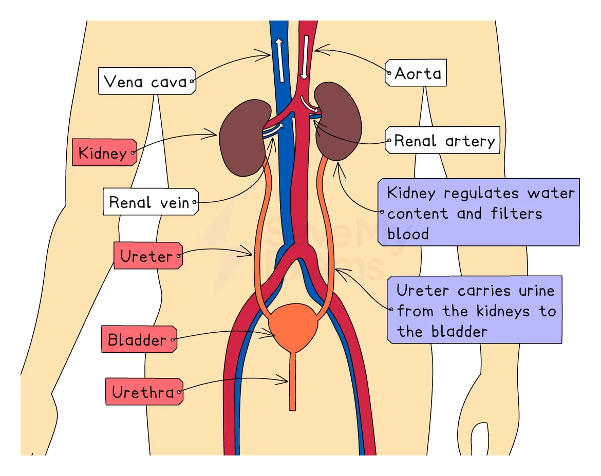
The kidneys are part of the urinary system, which filters the blood and removes water-soluble waste in the form of urine. The complete system has five named parts:
| Structure | What it does |
|---|---|
| Kidneys (one pair) | Filter the blood and form urine. Located on either side of the spine, towards the back of the abdomen, just below the ribs |
| Renal artery | Brings blood from the aorta into each kidney to be filtered. The renal artery has a high oxygen and a high urea content |
| Renal vein | Carries filtered blood away from each kidney back to the vena cava. The renal vein has a low urea content |
| Ureter (one per kidney) | The tube that carries urine from the kidney down to the bladder |
| Bladder | A muscular elastic bag that stores urine until it can be passed |
| Urethra | The single tube that carries urine from the bladder out of the body |
A tip for telling the two tubes apart: the ureter is one of two tubes that goes between upper organ (kidney) and bladder, while the urethra is the single tube that ends at the thigh.
4. Inside the kidney
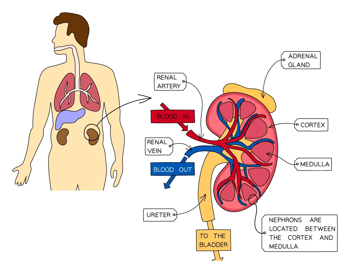
A vertical cross-section through a kidney shows three regions:
- Cortex is the outer layer. Contains the Bowman's capsules, glomeruli, proximal convoluted tubules and distal convoluted tubules (covered in section 5).
- Medulla is the inner darker layer. Contains the loops of Henle and most of the collecting ducts.
- Renal pelvis is the funnel-shaped cavity at the very centre. Urine drains into the renal pelvis from all the collecting ducts and flows from there into the ureter.
Each kidney contains roughly one million tiny filtering units called nephrons. Each nephron starts in the cortex, dips down into the medulla, and ends at the renal pelvis. Together they filter the entire blood plasma volume around 60 times every day.
5. The nephron
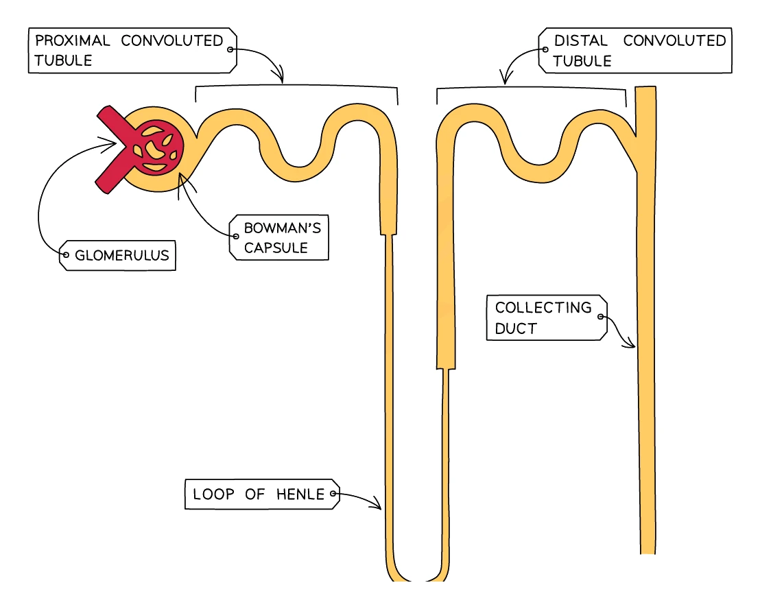
The nephron is the functional unit of the kidney: every step of urine formation happens along its length. A single nephron has these parts, in order along the route the filtrate takes:
- Bowman's capsule: a cup-shaped structure in the cortex that surrounds the glomerulus, a tight knot of capillaries. The Bowman's capsule is where blood is filtered (, section 6).
- Proximal convoluted tubule (PCT): a coiled tube also in the cortex. of glucose happens here.
- Loop of Henle: a long hairpin loop that dips deep down into the medulla and back up. Reabsorbs some water and salts.
- Distal convoluted tubule (DCT): a second coiled tube, back in the cortex. Fine-tunes the salt content of the filtrate.
- Collecting duct: runs back down through the medulla to the renal pelvis. The site of most water reabsorption, controlled by the hormone (section 7).
Around each nephron runs a dense network of capillaries that picks up the substances reabsorbed from the filtrate and returns them to the bloodstream.
Common exam question
Identifying structures on a kidney or nephron diagram
Question: Identify labelled structures on a diagram of the urinary system, a kidney section or a nephron, usually as one-mark multiple-choice items: the Bowman's capsule, loop of Henle or proximal convoluted tubule, where ultrafiltration happens, where glucose is reabsorbed, or which structure ADH affects (1 mark each).
Asked in 6 of the 23 papers. Know the nephron in filtrate order: the cup-shaped Bowman's capsule around the glomerulus (ultrafiltration); the first coiled tube, the proximal convoluted tubule (glucose reabsorbed); the hairpin loop of Henle in the medulla; the second coil, the distal convoluted tubule; then the collecting duct, which ADH acts on. On a whole-kidney section the renal artery and vein carry blood; only the ureter carries urine.
The ureter runs from kidney to bladder; the urethra leaves the bladder. One scheme rejects "urethra" for the ureter (a phonetic spelling that cannot be confused with it is allowed) and "gall bladder" for the bladder.
6. How urine is formed
Three main processes turn blood into urine as the filtrate flows along the nephron:
- in the glomerulus / Bowman's capsule (filtration under pressure)
- in the PCT (taking useful molecules back)
- Water reabsorption in the collecting duct (controlled by ADH)
Ultrafiltration
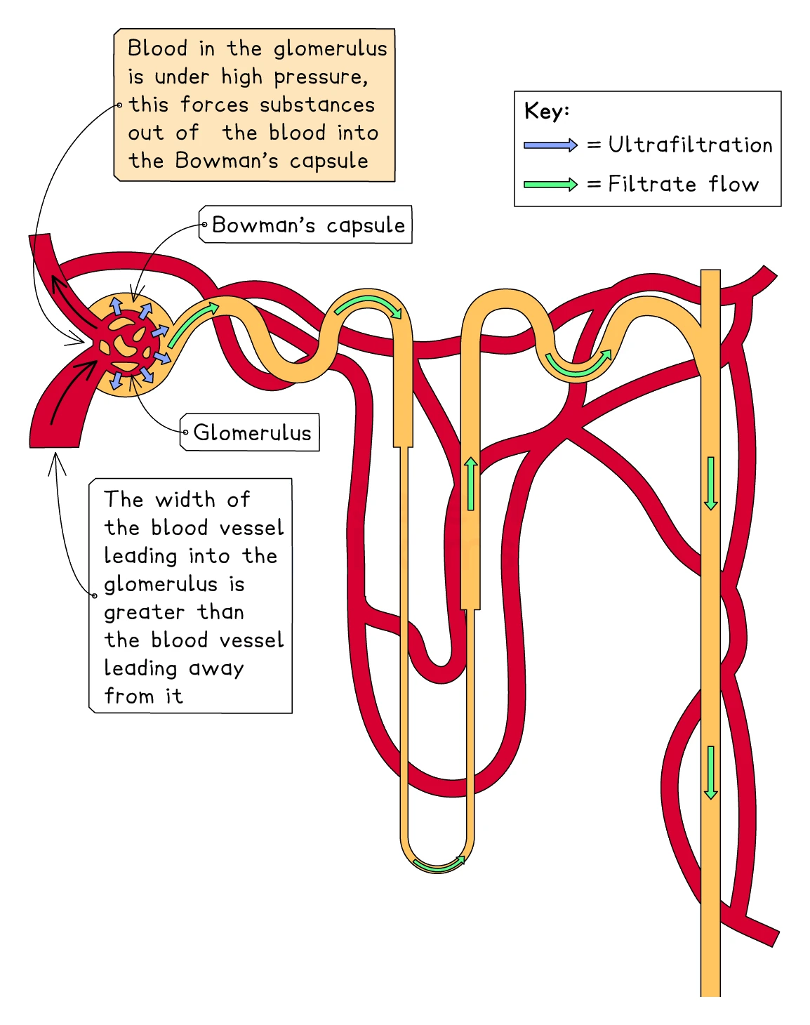
The Bowman's capsule surrounds a small knot of capillaries called the glomerulus. Two clever design features set up high-pressure filtration here:
- The afferent arteriole (the vessel bringing blood in) is wider than the efferent arteriole (the vessel taking blood out). This produces a bottleneck that raises the blood pressure inside the glomerulus to several times the normal capillary pressure.
- The wall of the glomerular capillary and the wall of the Bowman's capsule together form an extremely thin, partially-permeable filter.
The high blood pressure forces the small components of plasma out of the capillaries and across the filter into the Bowman's capsule:
- Water
- Glucose
- Amino acids
- Urea
- Mineral ions (Na⁺, K⁺, Cl⁻, etc.)
Larger molecules and cells are too big to cross the filter and stay in the blood:
- Red blood cells
- White blood cells
- Platelets
- Plasma proteins (such as albumin)
The fluid that collects inside the Bowman's capsule is called the glomerular filtrate. It is blood plasma without the proteins and cells.
High blood pressure in the glomerulus can damage the filter, allowing proteins to get through. Finding protein in urine is therefore an early sign of high blood pressure or kidney disease.
Common exam question
Explaining ultrafiltration and the absence of protein
Question: Explain why protein is not normally found in urine, why it appears in the urine of someone with high blood pressure, or why the vessel entering the glomerulus is wider than the one leaving it (2 marks).
Asked in 3 of the 23 papers. The protein marks are that protein molecules are large, so they cannot pass out of the glomerulus into the Bowman's capsule. High blood pressure forces protein through the filter, and once in the filtrate it is not reabsorbed. A longer question giving three patients' urine results wants the same point for the patient with protein present: ultrafiltration at the glomerulus is the process affected.
For the wide vessel in and narrow vessel out, any two of: the narrowing raises the pressure in the glomerulus; this is for ultrafiltration; so plasma, or small molecules such as urea, are forced into the Bowman's capsule. Say plasma or small molecules, not "blood", which is ignored.
Selective reabsorption in the PCT
The glomerular filtrate is full of valuable molecules that the body cannot afford to wash away. As the filtrate flows along the proximal convoluted tubule (PCT), these useful substances are pulled back into the bloodstream through the surrounding capillaries:
- All of the glucose is reabsorbed (by active transport, against its concentration gradient)
- All of the amino acids are reabsorbed
- Most of the mineral ions are reabsorbed
- A large amount of water is reabsorbed too, by osmosis (following the reabsorbed solutes)
It is selective because only some of the molecules are pulled back: useful ones move into the blood while waste substances (such as urea) are left behind in the filtrate.
Active transport of glucose against its gradient needs ATP, so the cells lining the PCT are packed with mitochondria. The wall of the PCT also has microvilli on its inner surface, dramatically increasing the surface area for reabsorption.
In a healthy person, all of the glucose in the filtrate is reabsorbed in the PCT, so there should be no glucose in the urine at all. If glucose is found in the urine, it usually means blood glucose has risen so high that the PCT cannot keep up. This is a classic sign of diabetes.
Common exam question
Explaining what happens to glucose in the kidney
Question: Explain what happens to glucose in the kidney, why the filtrate contains glucose but the urine does not, or why glucose is reabsorbed (2–3 marks).
Asked in 5 of the 23 papers. Two or three of these earn the marks, each a mark of its own in at least one scheme: glucose is filtered from the glomerulus into the Bowman's capsule; reabsorbed back into the blood; by active transport; using energy or ATP; against the concentration gradient; in the proximal convoluted tubule. Name the place as well as the process. Write "reabsorbed" or "absorbed back into the blood": one scheme ignores "absorbed" on its own.
If the question asks why glucose is reabsorbed, the points are purpose: so it is not lost from the body, to maintain blood glucose, and because it is needed for respiration. Glucose in the urine means the tubule has not taken it all back, the sign of diabetes.
Water reabsorption in the loop of Henle and collecting duct
After the PCT, the filtrate flows down through the loop of Henle into the medulla and back up. The loop of Henle uses a clever counter-current arrangement (beyond this level in detail) to make the surrounding tissue of the medulla very salty. This means that when the filtrate later flows through the collecting duct back down through the same salty tissue, water moves out of the filtrate by osmosis into the blood.
The amount of water that is reabsorbed here depends on how much the body needs to keep, and that is controlled by the hormone (section 7).
| Process | Where | What happens | Key substances |
|---|---|---|---|
| Ultrafiltration | Glomerulus → Bowman's capsule | High blood pressure forces small molecules through a partially-permeable filter | Water, glucose, urea, ions filtered out; proteins and cells stay in blood |
| Selective reabsorption | Proximal convoluted tubule | Useful molecules are reabsorbed by active transport into the blood | All glucose, all amino acids, most ions, much water |
| Water reabsorption | Collecting duct | Water moves into the blood by osmosis; amount controlled by ADH | Water |
What is left in the collecting duct at the end of all of this becomes urine: water + urea + excess ions + a few other waste products. The urine flows into the renal pelvis, down the ureter, into the bladder, and eventually out through the urethra.
Worked example
Calculations from a table of kidney data
The table gives data for the kidneys of a healthy adult over one day.
| Fluid | Volume per day in dm³ | Concentration of urea in g/dm³ |
|---|---|---|
| Glomerular filtrate | 180 | 0.3 |
| Urine | 1.8 | 20 |
(a) Calculate the percentage of the filtrate volume that is reabsorbed.
(b) Work out how many times greater the urea concentration is in the urine than in the filtrate, to two significant figures.
Solution:
- (a) Volume reabsorbed = 180 − 1.8 = 178.2 dm³
- Percentage reabsorbed = 178.2 ÷ 180 × 100 = 99%
- (b) Concentration factor = 20 ÷ 0.3 = 66.67…
- To two significant figures, the urea is 67 times more concentrated in the urine
Write the division or the subtraction down before you reach for the calculator. In the real questions a correct answer alone earns both marks, but when the arithmetic slips, the written step still earns one of them.
7. ADH and osmoregulation
Osmoregulation is the control of the water content and salt concentration of the body fluids within narrow limits
Exam tip
Define osmoregulation with salt as well as water
When a question asks what osmoregulation means (2 marks), "controlling the water content of the body" is only half the definition and earns one mark. The second mark needs the dissolved substances too: osmoregulation maintains the water and salt (solute) concentration, or the water potential, of the blood and body fluids. Give both halves in one sentence.
Body cells only function properly within a small range of water content. Too little water in the blood and cells lose water by osmosis and shrink. Too much water and cells swell and may burst. The kidneys keep things in balance by adjusting how much water they leave in the urine.
The hormone that controls this adjustment is (antidiuretic hormone). The word "antidiuretic" means "against urine production", so high ADH makes the kidneys produce less urine.
How ADH works (negative feedback)
The body uses a negative feedback loop to keep blood water content steady. The loop has these stages:
- Detection. Special cells called osmoreceptors in the hypothalamus of the brain monitor the water concentration of the blood as it flows past.
- Signalling. The hypothalamus tells the pituitary gland (just beneath it) to release more or less ADH into the blood.
- Effect on the kidney. ADH circulates to the kidneys, where it changes how permeable to water the walls of the collecting ducts are.
- Reabsorption. With more permeable walls, more water moves out of the filtrate back into the blood, leaving less in the urine. With less permeable walls, less water is reabsorbed and more ends up in the urine.
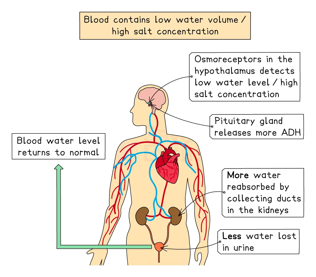
When blood water is too LOW (dehydrated)
For example, after exercise on a hot day, or after eating a salty meal:
- Osmoreceptors detect that the blood is too concentrated (low water).
- Hypothalamus signals the pituitary to release more ADH.
- ADH makes the collecting ducts more permeable to water.
- More water is reabsorbed from the filtrate back into the blood.
- The body produces a small volume of concentrated, dark yellow urine.
- Blood water rises back to normal. (You also feel thirsty and drink water, which adds more.)
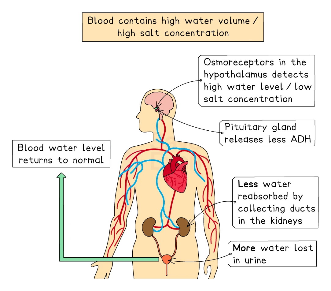
When blood water is too HIGH (over-hydrated)
For example, after drinking a litre of water:
- Osmoreceptors detect that the blood is too dilute (high water).
- Hypothalamus signals the pituitary to release less ADH.
- ADH levels drop. Collecting ducts become less permeable to water.
- Less water is reabsorbed; more passes into the urine.
- The body produces a large volume of dilute, pale yellow urine.
- Blood water falls back to normal.
Key mark-scheme phrase: "ADH increases the permeability of the collecting duct to water." Exam mark schemes expect this exact wording.
Common exam question
Explaining the ADH response to dehydration
Question: Explain the changes in the body when a person is dehydrated (after exercise, a salty meal, or vomiting and diarrhoea) that lead to a small volume of concentrated urine (3–5 marks).
Asked in 6 of the 23 papers. The marks follow the chain, usually one per link: the water content of the blood falls; osmoreceptors in the hypothalamus detect this; the pituitary releases more ADH; ADH makes the collecting duct more permeable to water; so more water is reabsorbed into the blood and less, more concentrated urine is made. Name the collecting duct and say what changes about it: one scheme marks each separately.
If the question compares pure water with an isotonic drink, reverse the chain: blood more dilute, less ADH, less water reabsorbed, so more, paler urine; the isotonic drink's salt and sugar keep the blood at normal concentration. A two-mark describe version wants only the urine: less of it, and more concentrated or darker.
8. The composition of urine
In a healthy person, urine is roughly:
| Substance | What it is | Where it came from |
|---|---|---|
| Water (about 95%) | Solvent | Filtrate that was not reabsorbed |
| Urea | The body's main nitrogen-containing waste | Made in the liver from excess amino acids |
| Excess ions | Mostly sodium, potassium and chloride | Filtered from blood; only the excess is excreted |
| Other small wastes | Creatinine, broken-down drug molecules, hormone breakdown products | Various metabolic reactions |
Urine should not normally contain:
- Glucose (all reabsorbed in the PCT)
- Amino acids (all reabsorbed)
- Proteins (too large to be filtered)
- Blood cells (too large to be filtered)
Finding any of these in urine is a sign of disease. Glucose suggests diabetes; protein suggests kidney damage or very high blood pressure.
Common exam question
Testing a urine sample for protein or glucose
Question: Describe how a sample of urine could be tested for protein, or for glucose (2 marks).
Asked in 4 of the 23 papers, twice for each substance. For protein, add biuret reagent (or sodium hydroxide then copper sulfate) and look for a lilac, purple or pink colour: one mark for the reagent, one for the colour. For glucose, add Benedict's solution and heat; a green, yellow, orange or red colour shows glucose is present. Give all three: one scheme credits any two of the reagent, the heating and the colour, but the other folds the heating into the reagent mark, so only the colour earns the second.
Test strips are accepted in all four schemes, and one scheme also accepts ninhydrin (going pink or red) for protein. Whichever reagent you name, give the colour that belongs to it: the strip's own colour change, brown for a glucose strip, is what earns the colour mark.
What changes the colour and volume of urine
The volume and concentration of urine can change rapidly:
- More fluid drunk = larger volume of paler urine
- Hot weather or vigorous exercise = more water lost as sweat, so a smaller volume of darker urine
- Cold weather = less sweating, so more urine
- Salty diet = body needs to lose excess salt, often as concentrated urine
- Caffeine and alcohol = both block or reduce ADH release, producing larger volumes of paler urine and contributing to dehydration
9. Kidney failure
Each kidney has so much spare filtration capacity that a person can live a normal life with only one working kidney. But if both kidneys fail (a condition called renal failure), urea, water and ions accumulate in the blood within days, and the patient will die without treatment. There are two main treatments: and kidney transplant.
Dialysis (an artificial kidney)
Dialysis is a machine-based treatment that artificially performs the kidney's filtration job. The most common type is haemodialysis:
- Blood from one of the patient's arteries flows out of the body and into the dialysis machine.
- Inside the machine, the blood passes through a long network of partially permeable tubes (the artificial filter).
- Outside these tubes flows a dialysis fluid designed to match the normal levels of glucose, mineral ions and water that should be in healthy blood. The fluid contains no .
- Urea, excess ions and excess water diffuse out of the blood through the partially permeable membrane into the dialysis fluid (down a concentration gradient).
- Useful substances such as glucose and the right amount of ions stay in the blood, because the dialysis fluid already has the same concentration of these (no gradient → no net diffusion).
- The cleaned blood returns to a vein, back into the body.
A typical haemodialysis session lasts about 4 hours and is done three times a week at a hospital or clinic. Patients have to:
- Watch their diet closely (especially salt and protein intake, since the machine is not as efficient as a real kidney at controlling these)
- Limit how much fluid they drink between sessions
- Accept the time commitment, the restrictions on travel, and the long-term risks of infection at the access point
Common exam question
Explaining how dialysis removes urea but keeps glucose
Question: Explain how the composition of the dialysis fluid removes urea but keeps glucose and ions in the blood, or how the dialyser is designed to speed up the removal of urea (2–4 marks).
Asked in 2 of the 23 papers. Urea leaves because the fluid contains no urea (a lower concentration of urea, excess salts and other waste), so there is a concentration gradient and urea diffuses out of the blood across the partially permeable membrane. Glucose and ions stay because the fluid holds them at the same concentration as plasma: no gradient, no net loss, and the body needs glucose for respiration and water and ions for water balance.
The design question marks in pairs, a feature with its reason, so give both: fluid kept moving to maintain the gradient; long, coiled tubing for a large surface area; a thin membrane for a short diffusion distance; fluid kept warm so particles move faster.
Dialysis keeps people alive, sometimes for many years, but it is not a cure. Without it, kidney failure is fatal within days.
Kidney transplant
A kidney transplant replaces the failed kidney with a healthy one from a donor. The donor can be:
- A close living relative (who can donate one kidney and survive normally with the other)
- A recently deceased person whose family has consented to organ donation
The surgery places the donor kidney into the lower abdomen, connects its blood vessels to the patient's arteries and veins, and connects its ureter to the patient's bladder. If everything works, the patient's body fluid balance returns to normal within days.
Advantages of a transplant over :
- A working transplanted kidney is far more efficient than a dialysis machine
- The patient does not need to attend the clinic three times a week
- Diet and fluid restrictions can usually be relaxed
- The cost per year is much lower in the long run
Disadvantages and challenges of a transplant:
- The donor kidney is recognised by the recipient's immune system as foreign because its antigens do not match. The recipient's lymphocytes can attack the transplant, a process called rejection.
- To reduce the chance of rejection, the donor must be a tissue match (the donor's antigens must be as close to the recipient's as possible). Close relatives are usually the best matches.
- The recipient must take immunosuppressant drugs for life to dampen the immune response. This makes them more vulnerable to infections and some cancers.
- There is a shortage of donor kidneys, and the waiting list is often long. Many patients spend years on dialysis before a suitable kidney becomes available.
Exam tip
Transplant questions: tissue match and credited advantages
Asked why a kidney from a close relative is less likely to be rejected (2 marks), give two of: the relative is genetically similar; so the kidney has the same or similar antigens, tissue type or blood type; so there is less immune response and fewer antibodies. Asked for advantages of a transplant over dialysis, credited points are that it is permanent, allows a normal life and diet with no repeated hospital visits, and that the kidney does its other functions. Repeating a reason the passage already gave earns nothing, and "cheaper in the long run" is ignored.
| Dialysis | Kidney transplant | |
|---|---|---|
| What it does | Filters the blood with an external machine | Replaces the failed kidney with a working donor kidney |
| How often | Three sessions of around 4 hours per week, for life | One surgery; the new kidney then works continuously |
| Lifestyle restrictions | Strict diet and fluid limits, frequent hospital visits | Relatively normal life; daily immunosuppressant tablets |
| Donor needed? | No | Yes (living relative or deceased donor) |
| Risk of rejection | None (no donor tissue involved) | Yes; must be tissue-matched and take immunosuppressants |
| Long-term cost | High (machine time, staff time, transport) | Lower in the long run, once surgery is paid for |
| Quality of life | Substantially reduced | Close to normal if the kidney is not rejected |