1. Detecting change and responding to it
Every living organism has to detect what is happening around it and respond in a way that keeps it alive. A change in the environment that an organism can detect is called a stimulus; the action the organism then takes is called the response.
Every coordinated response uses the same five-step pathway:
stimulus → receptor → coordinator → → response
| Stage | What it is | Examples |
|---|---|---|
| Stimulus | A change the organism can detect | Light, sound, temperature, touch, pressure, chemical signals |
| Receptor | A cell or organ that detects the stimulus | Light-sensitive cells in the eye; touch receptors in the skin; taste buds; the inner ear |
| Coordinator | A control centre that processes information and decides what to do | Brain, spinal cord, pancreas |
| Effector | A muscle or gland that carries out the response | Skeletal muscle (contracts); sweat gland (secretes sweat); endocrine gland (releases a hormone) |
| Response | The action taken | Pulling a hand away, sweating to cool down, releasing insulin |
In humans, two systems carry out this coordination: the nervous system (fast, electrical signals) and the endocrine system (slower, chemical signals). They work side by side, each one good for different jobs.
2. Nervous coordination vs hormonal coordination
The two systems differ in speed, signal type, target, and duration of effect.
| Feature | Nervous system | Endocrine (hormonal) system |
|---|---|---|
| Signal type | Electrical impulses along neurones | Chemical signals (hormones) carried in the blood |
| Pathway | Through nerves (bundles of neurones) | Through the bloodstream |
| Speed | Very fast (impulses up to 100 m/s) | Slower (seconds to hours) |
| Target | Specific cells at the end of the neurone | Many cells, but only those that have the matching receptors respond |
| Duration of response | Short (the response stops the moment the signal stops) | Long (the response lasts until the hormone is broken down) |
| Example | The pain-withdrawal reflex | Insulin lowering blood glucose; adrenaline raising heart rate |
The nervous system is the right tool for jobs that need an immediate response: pulling away from a sharp object, flinching from a loud noise, balancing on a moving bus. The endocrine system is the right tool for longer-lasting changes that affect lots of cells at once: growing taller, going through puberty, regulating blood sugar between meals.
Common exam question
Differences between nervous and hormonal communication
Question: Describe three differences between the nervous system and the hormonal system, or between hormones and neurotransmitters (3 marks).
Asked in 2 of the 23 papers, one mark per difference. The credited pairs: an impulse travels along neurones, a hormone in the blood plasma; the nerve signal is electrical, the hormone chemical; a neurone (or neurotransmitter) acts on specific target cells, a hormone on many cells all round the body; a nervous response is short-lived, a hormonal one longer-lasting. For hormones against neurotransmitters, where each is made (endocrine gland, end of a neurone) is a fourth pair. Either side of a pair earns its mark.
Speed is the difference everyone knows, yet neither scheme credits it: one paper used it in a ratio first and asks for "other" differences; the other ignores faster or slower. In that ratio, converting to the same units (×60 or ÷60) earns a mark, dividing another, the form n : 1 the third.
3. The human nervous system
The human nervous system is built from two parts:
- The central nervous system (CNS): the brain and spinal cord. This is where the decisions are made.
- The peripheral nervous system (PNS): every nerve outside the CNS. These connect the CNS to the receptors and effectors all over the body.
A nerve is a bundle of long thin cells called neurones. Each neurone carries electrical impulses along its length.
Three types of neurone
| Neurone | Carries impulses... | Found in... |
|---|---|---|
| Sensory neurone | From a receptor to the CNS | The PNS |
| Relay neurone | Within the CNS, from one neurone to another | The CNS only |
| Motor neurone | From the CNS to an effector | The PNS |
The standard pathway through the nervous system, in summary form:
receptor → sensory neurone → (relay neurones) → motor neurone →
4. Synapses: passing the signal between neurones
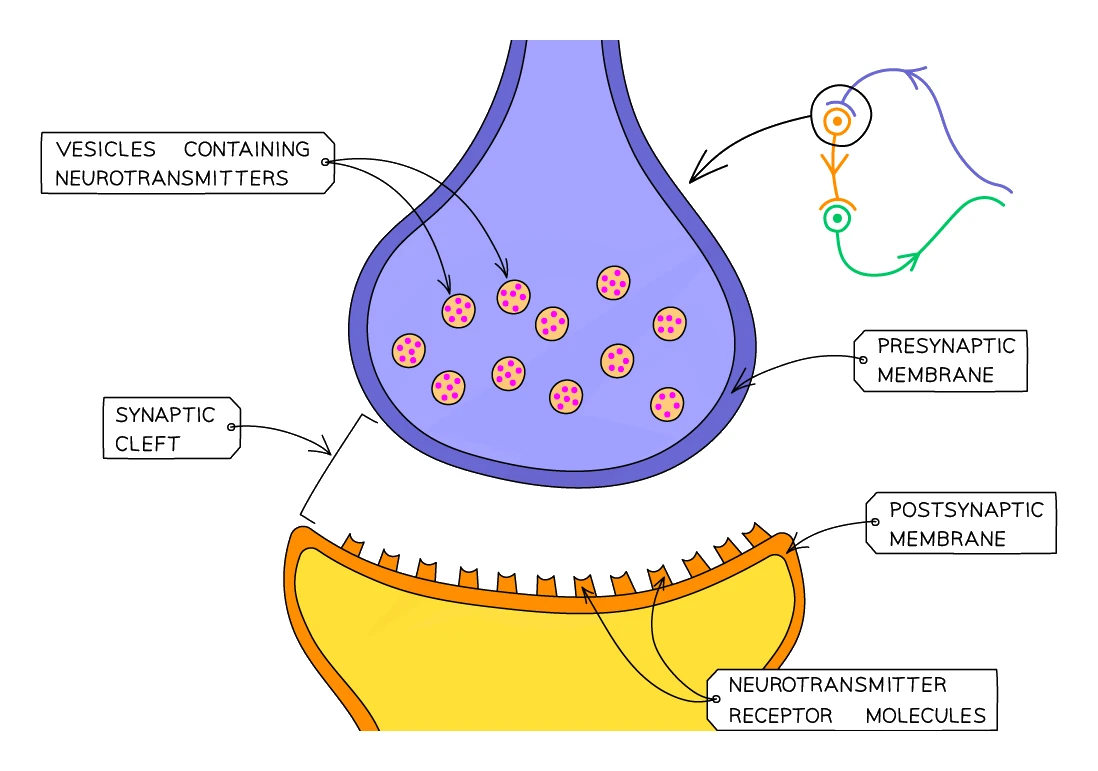
Neurones are not actually connected to each other. A tiny gap, called a , sits between the end of one neurone and the start of the next. The electrical impulse cannot jump across this gap; instead, the message has to be turned into a chemical signal for the crossing.
How a synapse works
- An electrical impulse travels along the first neurone and arrives at the end of it (called the presynaptic terminal).
- The arrival of the impulse triggers the release of neurotransmitter molecules stored in tiny sacs in the end of the neurone. The neurotransmitters spill out into the synaptic gap.
- The neurotransmitters diffuse across the gap to the next neurone.
- They bind to receptor proteins on the membrane of the next neurone.
- This triggers a new electrical impulse in the next neurone, which then carries the signal onwards.
- The neurotransmitter is quickly broken down or reabsorbed so the signal does not last forever.
Why synapses are useful
- One-way only. Neurotransmitter is released only on one side of the synapse and detected only on the other, so signals can only pass in one direction. This stops signals bouncing back and getting tangled.
- Signal modulation. Synapses can amplify, dampen or block a signal. Many drugs (and some natural processes) work by changing what happens at a synapse.
Common exam question
How an impulse crosses a synapse
Question: Describe how an impulse passes from one neurone to the next, or explain why the time taken to respond differs from the time an impulse needs to travel along the neurones (2–4 marks).
Asked in 2 of the 23 papers. The two-mark version wants any two of: the gap is a synapse; a neurotransmitter is released and travels across it; it crosses by diffusion (chemical, not electrical, coordination).
The response-time version is the same mechanism in disguise; four marks from: the response takes longer than the travel time; there is a delay between neurones, not just travel along them; that delay is at the synapse, crossed by a neurotransmitter by diffusion; and time to generate the impulse in the receptor and for the muscle to contract. The phrase "synapses between neurones" collects two of those points at once, and "the impulse travels faster" is ignored. Synapse and neurotransmitter also earn marks inside a reflex-arc description.
5. Reflex actions and the reflex arc
A reflex action is a very fast, automatic, involuntary response to a stimulus. You do not have to think about it; by the time you become aware of the stimulus, your body has already responded. Examples include pulling your hand away from a sharp object, blinking when something flies near your eye, coughing when food goes the wrong way, and the pupil narrowing in bright light.
Reflex actions are useful because they are fast and automatic, which protects the body from damage in dangerous situations.
The reflex arc
The neural pathway followed by a reflex action is called a . It bypasses the conscious part of the brain to save precious milliseconds. The arc usually goes through the spinal cord rather than the brain.
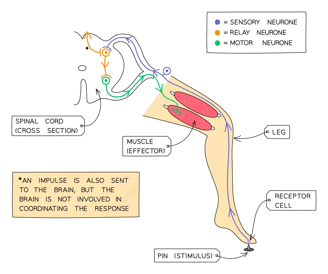
Tracing the reflex when a person steps on a sharp pin:
- Stimulus: the sharp point of the pin pressing on the foot
- Receptor: pain receptors in the skin of the foot detect the pressure and damage
- Sensory neurone: an electrical impulse travels from the receptor up the leg into the spinal cord
- Relay neurone: inside the spinal cord, the sensory neurone synapses with a short relay neurone, which immediately synapses with a motor neurone. This is the arc: the signal arcs across the spinal cord without going up to the brain
- Motor neurone: an impulse travels back down the leg to the muscles
- Effector: the muscles in the leg contract
- Response: the foot is pulled up and away from the pin
The whole thing takes a fraction of a second. A separate, slower signal also goes up to the brain so that you become aware of the pain shortly after the foot has moved.
The reflex arc: stimulus → receptor → sensory neurone → relay neurone (in spinal cord) → motor neurone → → response
Common exam question
Describing the pathway of a reflex arc
Question: Describe the pathway of a nerve impulse in a withdrawal reflex arc, or identify its receptor, neurones and effector on a diagram (4 marks, or 1 mark per structure).
Set in 4 of the 23 papers. Four marks for the description: the receptor detects the stimulus and generates an impulse; along the sensory neurone to the spinal cord; across a synapse (by neurotransmitter) to the relay neurone, then the motor neurone; to the effector, the muscle, which contracts. Name the sensory and motor neurones, each a separate point, and write impulse: "signal" and "message" are ignored.
On a diagram, the receptor is where the stimulus is detected, the relay neurone's cell body is inside the spinal cord, the motor neurone runs from the cord to the muscle (the effector), and an impulse arrow on the motor neurone points away from the cord. Asked what a reflex is, or its name: automatic, involuntary, done without thinking, a withdrawal reflex.
Worked example
Time for an impulse to travel along a reflex arc
The nerve pathway of a withdrawal reflex is 140 cm long and the impulse travels along it at 80 metres per second. Calculate the time taken for the impulse to travel the whole pathway. Give your answer in standard form.
Solution:
- Convert the distance to metres so the units match the speed: 140 cm = 1.40 m
- time = distance ÷ speed = 1.40 ÷ 80 = 0.0175 s
- In standard form: 1.75 × 10⁻² s
This calculation is set in 2 of the 23 papers, for 2 marks. A correct answer alone earns both. In one scheme, dividing the distance by the speed earns one mark even when the answer is wrong; the other paper demanded standard form and gave only one mark to the right value written as an ordinary decimal, so check the form asked for. The real response takes longer than this, because of the synapses described above.
6. The human eye
The eye is a sense organ that detects the stimulus of light. It contains light-sensitive receptor cells, plus a sophisticated lens system that focuses light onto those cells.
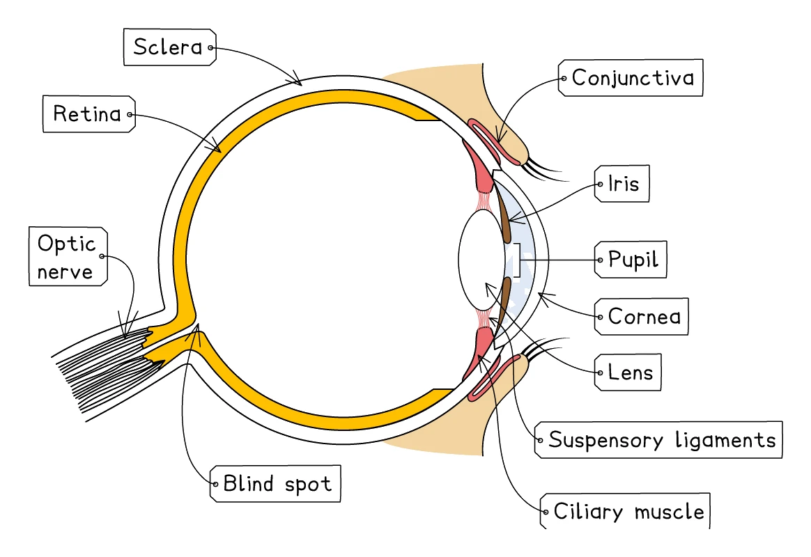
The main structures of the eye
| Structure | What it does |
|---|---|
| Cornea | A see-through, dome-shaped tissue at the very front of the eyeball. Refracts (bends) light as it enters, doing most of the focusing |
| Sclera | The tough white outer wall of the eyeball. Keeps the eye's shape; provides attachment for the muscles that move the eye |
| Conjunctiva | A thin transparent membrane covering the front of the eye; keeps it moist and protected |
| Iris | The coloured ring of muscle that controls how much light enters by changing the size of the pupil |
| Pupil | The black hole in the centre of the iris; the opening that lets light in (the pupil itself is just space; it looks black because the inside of the eye is dark) |
| Lens | A clear flexible disc that fine-focuses the light onto the retina. Can change shape to focus on near or far objects |
| Ciliary muscle | A ring of muscle that surrounds the lens. Changing its tension changes the shape of the lens |
| Suspensory ligaments | Strong fibres that connect the lens to the ciliary muscle |
| Retina | The light-sensitive layer at the back of the eye. Contains rod cells (for dim light, no colour) and cone cells (for colour) |
| Fovea | A patch of retina directly opposite the lens, packed with cone cells. Gives the sharpest, most detailed colour vision |
| Optic nerve | Carries impulses from the retina to the brain |
| Blind spot | The point where the optic nerve leaves the retina. No receptor cells here, so any image falling on this spot is not seen |
Common exam question
Identifying the parts of the eye on a diagram
Question: On a labelled section through the eye, name a structure, or choose which structures refract light, which controls the light reaching the retina and which holds the light-sensitive cells (1 mark each).
Set in 3 of the 23 papers. The cornea and the lens both refract light onto the retina; the iris controls how much light reaches it; the retina holds the light-sensitive cells; the coloured ring round the pupil is the iris. Know the diagram well enough to tell retina, conjunctiva and lens apart when all three are offered.
The question can move on to a cataract, a cloudy or opaque lens: say that less light passes through it (refraction is affected) to the retina's photoreceptors, so fewer impulses reach the brain and the image is dim or blurred. "Poor vision" alone is ignored. Once the cloudy lens is removed, the person needs glasses, contact lenses or an artificial lens; laser surgery is ignored.
Accommodation: focusing on near and far objects
The eye uses two parts to focus light: the cornea does most of the bending, and the lens does the fine-tuning. The lens can change shape, which lets the eye focus on objects at different distances. This is called .
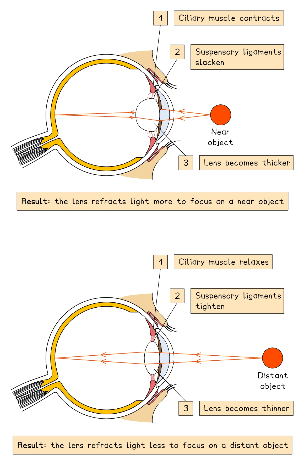
Looking at a near object (e.g. reading a book):
- The ciliary muscle contracts, so the ring of muscle becomes smaller in diameter
- This slackens the suspensory ligaments
- The lens, no longer pulled tight, becomes fatter and more rounded
- A fatter lens refracts (bends) the light more, focusing the light from the close object onto the retina
Looking at a far object (e.g. a tree on a distant hill):
- The ciliary muscle relaxes, so the ring of muscle becomes wider in diameter
- This pulls the suspensory ligaments tight
- The taut ligaments pull on the lens, making it thinner and flatter
- A thinner lens refracts the light less, which is enough for the parallel rays of light from the distant object to focus on the retina
A common mistake: people sometimes write that "the suspensory ligaments contract". They don't, because they are ligaments, not muscles. The right words are tighten or slacken.
| Near object | Distant object | |
|---|---|---|
| Ciliary muscle | Contracts | Relaxes |
| Suspensory ligaments | Slack | Taut |
| Lens shape | Fat / rounded | Thin / flat |
| Light refraction | More | Less |
Common exam question
Focusing on a near object
Question: Describe the changes in the eye, or how the ciliary muscle and the suspensory ligaments control the shape of the lens, when it focuses on a near object (3–4 marks).
Asked in 2 of the 23 papers, one mark per step, so give the whole chain in order: the ciliary muscles contract; the suspensory ligaments slacken, their tension reduced; the lens becomes thicker, fatter, more rounded or more convex; so it refracts light more. Naming the process, accommodation, was a fifth point in one scheme.
Both schemes ignore "the suspensory ligaments relax": ligaments are not muscles, so write slacken, loosen or lose tension, and never call them a suspensory muscle. When the two structures are lettered on a diagram, say which letter is the ciliary muscle and which the ligaments before describing what each does.
The pupil reflex: responding to light intensity
The amount of light reaching the retina is controlled by the size of the pupil. The iris changes the pupil size automatically through a reflex, protecting the retina from too much light and helping with vision in dim light.
The iris contains two sets of muscles, working as antagonistic pairs:
- Circular muscles run around the pupil. When they contract, the pupil constricts (narrows).
- Radial muscles run from the centre outwards like spokes on a wheel. When they contract, the pupil dilates (widens).
In bright light:
- Photoreceptors detect high light intensity
- The circular muscles contract, the radial muscles relax
- The pupil constricts (becomes smaller)
- Less light enters the eye, protecting the retina from damage
In dim light:
- Photoreceptors detect low light intensity
- The circular muscles relax, the radial muscles contract
- The pupil dilates (becomes larger)
- More light enters the eye, improving vision
| Bright light | Dim light | |
|---|---|---|
| Circular muscles | Contract | Relax |
| Radial muscles | Relax | Contract |
| Pupil size | Narrow | Wide |
| Light entering | Less | More |
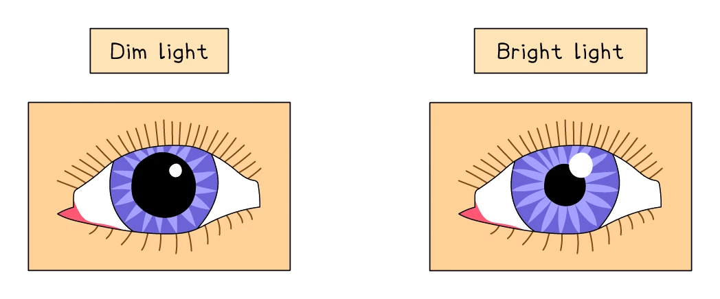
The pupil reflex is automatic; you do not consciously control it. It is a classic example of a reflex action coordinated by the brain (specifically, by the brain stem) rather than by the spinal cord.
Common exam question
How the iris controls the light entering the eye
Question: Explain the changes in the iris when a person moves from dim light into bright light, or how an iris that lets light pass through it affects vision (2–3 marks).
Asked in 2 of the 23 papers. Moving into bright light, the circular muscles contract and the radial muscles relax, so the pupil constricts and less light reaches the retina. Any two of those three points score, so name both sets of muscles and say what the pupil does. Write circular, not ciliary: the ciliary muscle changes the lens, and the scheme ignores it here.
An iris with no pigment, as in a pink-eyed albino animal, does not block light, so changing the pupil has little effect and the iris cannot regulate the light entering the eye; too much light falls on the retina, giving poor vision or damaging it. "The iris lets light through" repeats the question and does not score on its own.
7. The skin in temperature regulation
Keeping the body's core temperature close to 37 °C is essential because the body's enzymes only work properly within a narrow temperature range. Even a 2 °C swing is potentially fatal. The skin plays a major role in adjusting heat loss to keep core temperature steady.
Temperature receptors in the skin and in the hypothalamus (a region at the base of the brain) detect changes in body temperature. The hypothalamus then coordinates the response, which is carried out by structures in the skin.
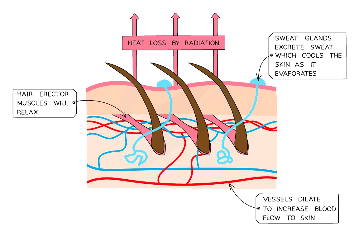
When the body is too hot
Three cooling mechanisms in the skin:
- of skin arterioles. The small arterioles that supply the surface capillaries widen (because the muscles in their walls relax). This sends more blood close to the skin's surface, where it loses heat to the surrounding air by radiation. The skin looks flushed red.
- Sweating. Sweat glands secrete more sweat onto the skin's surface. Water in the sweat evaporates, taking thermal energy from the body with it as latent heat. The skin cools as it evaporates.
- Hairs lie flat. Hair erector muscles relax, so the hairs lie flat against the skin. Less air is trapped close to the skin, so the insulating layer is thinner and heat can escape more easily.
When the body is too cold
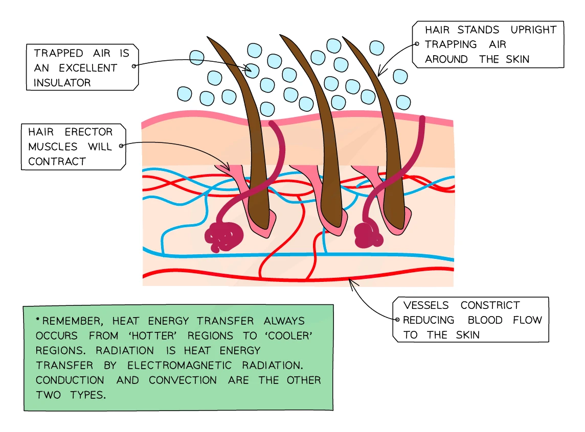
Three warming (or heat-conserving) mechanisms:
- of skin arterioles. The arterioles narrow (their muscles contract), sending less blood through the surface capillaries. Less heat is lost by radiation. The skin looks pale.
- Less sweating. Sweat glands secrete less sweat, so less heat is lost by evaporation.
- Shivering. Skeletal muscles contract and relax rapidly. The contractions need ATP, which is supplied by respiration; respiration releases heat as a by-product, warming the body.
- Hairs stand on end. Hair erector muscles contract, pulling the hairs upright. This traps a layer of air next to the skin. Air is a poor conductor of heat, so the trapped air acts as insulation, reducing heat loss. (We have less hair than most mammals, so this gives "goosebumps" but does not insulate much in humans.)
Note for exams: the Edexcel mark scheme expects students to mention sweating, vasodilation and vasoconstriction by name. Hair erection (goosebumps) is mentioned in the textbook but is not strictly required.
Common exam question
How the skin responds when the body is too hot or too cold
Question: Explain how the skin's blood vessels regulate body temperature when it rises, or describe how the skin responds to a cold environment (3–4 marks).
Asked in 3 of the 23 papers. Too hot: vasodilation; the arterioles supplying the skin capillaries widen; more blood flows near the surface; more heat is lost by radiation. Too cold, four marks from: vasoconstriction; the arterioles narrow ("arterioles constrict" scores both of these at once); less blood reaches the surface; the hair erector muscles contract so the hairs stand up; trapping air, which insulates; less heat is lost by radiation; less sweat is made.
The schemes police the words: capillaries have no muscle, so "capillaries dilate", "vasodilation of the capillaries" and "veins widen" are rejected, as is any idea that blood vessels move towards or away from the surface. Sweating is ignored when the question is about the blood vessels, and shivering is ignored in the cold-skin question: that is muscle, not skin.
8. Major human hormones
A hormone is a chemical messenger produced by an endocrine gland and carried in the blood to target cells in another part of the body. Endocrine glands have no ducts; they secrete their hormones directly into the bloodstream.
The major endocrine glands and their key hormones are summarised below. The detailed mechanisms (such as how insulin lowers blood glucose, or how ADH controls water reabsorption) are covered in topic 11 Homeostasis and topic 9 Excretion respectively.
| Hormone | Where it is made | Main role |
|---|---|---|
| Adrenaline | Adrenal glands (one above each kidney) | "Fight-or-flight" response: prepares the body for sudden physical action. Raises heart rate, raises breathing rate, redirects blood to muscles, breaks down stored glycogen in the liver to release glucose |
| Insulin | Pancreas | Lowers blood glucose. Stimulates muscles and liver to take in glucose from the blood and store it as glycogen |
| Testosterone | Testes (in males) | Triggers male puberty: development of male reproductive organs, growth of facial/body hair, deepening of the voice, increased muscle mass |
| Oestrogen | Ovaries (in females) | Triggers female puberty: breast development, widening of the hips, body hair. Also rebuilds the uterus lining after each menstrual period |
| Progesterone | Ovaries | Maintains the thick uterus lining ready for a fertilised egg, and during pregnancy. Prevents further ovulation while a fertilised egg is implanted |
| ADH (antidiuretic hormone) | Pituitary gland (just under the brain) | Increases the permeability of the kidney's collecting ducts to water, allowing the body to reabsorb more water and produce less urine (see topic 9) |
| FSH (follicle-stimulating hormone) | Pituitary gland | Causes an egg to mature in the ovary each month; stimulates the ovary to release oestrogen |
| LH (luteinising hormone) | Pituitary gland | Triggers ovulation (the release of the mature egg); stimulates the ovary to release progesterone |
Common exam question
Naming the gland, target and effect of a hormone
Question: Name or pick out the gland that produces a hormone, or complete a table of hormone, gland, target and effect (1 mark per answer).
Set in 6 of the 23 papers. The pairings the schemes credit: insulin from the pancreas, turning blood glucose into glycogen; adrenaline from the adrenal glands, raising heart rate; testosterone from the testes, driving male secondary sexual characteristics such as body hair (its target is the skin, not the testes); progesterone from the ovary, maintaining the uterus lining; FSH and LH from the pituitary gland, acting on the ovaries: FSH grows a follicle, LH triggers ovulation. "Pituitary" is enough; anterior or posterior adds nothing.
On a body outline, the pituitary sits beneath the brain, the adrenal glands on the kidneys, the pancreas below the stomach, the ovaries low in the female abdomen and the testes outside the male body. Fill every gap: one table gave a mark only for each fully correct row.
How adrenaline works (the "fight-or-flight" response)
The adrenal glands release adrenaline in response to fear, excitement, or sudden physical demand. Adrenaline causes a coordinated set of changes that prepare the body to either fight a threat or run from it:
- Heart rate increases. Blood is pumped to the muscles faster.
- Breathing rate and depth increase. More oxygen enters the blood; more CO₂ leaves.
- Pupils dilate. More light enters the eye, improving vision in low-light or distant scenes.
- Blood is redirected away from non-essential organs (such as the gut) and towards muscles. The arterioles supplying muscles dilate; those supplying the gut constrict.
- Glucose is released into the blood. Adrenaline triggers the liver to break down stored glycogen back into glucose, raising blood sugar so muscle cells have more fuel for respiration.
All of these effects begin within seconds of the adrenaline reaching its target cells and fade within minutes once the danger has passed.
Common exam question
Where adrenaline is made and what it does
Question: Describe where adrenaline is produced and its effects on the body, or explain how a change in adrenaline production would affect an animal's behaviour (3–4 marks).
Asked in 2 of the 23 papers. The credited points, one mark each: made by the adrenal glands; heart rate increases; the body is prepared for fight or flight; more blood flows to the muscles, diverted from the intestines; breathing rate and depth increase; glycogen is converted to glucose, raising blood glucose; pupils dilate; reactions are faster. Four marks need four of these, so go well beyond heart rate.
For the behaviour version, link the chain: larger adrenal glands make more adrenaline, which raises heart rate and the fight-or-flight response, so the animal shows quicker reflexes and reactions; more neurotransmitter likewise changes what happens at the synapses. The converse chain for smaller glands and less neurotransmitter is accepted too.
9. Plant responses: tropisms
Plants cannot move from place to place, but they can change their orientation by growing one way rather than another. A directional growth response in a plant is called a tropism.
A tropism is named after the stimulus it responds to:
- Phototropism is a response to light. Shoots show positive (grow towards the light); roots show negative phototropism (grow away from light).
- Geotropism (also called gravitropism) is a response to gravity. Roots show positive geotropism (grow downwards, in the direction of gravity, anchoring the plant and reaching water); shoots show negative geotropism (grow upwards, against gravity, lifting the leaves towards the sky).
| Plant part | Phototropism | Geotropism |
|---|---|---|
| Shoots | Positive (towards light) | Negative (away from gravity, i.e. upwards) |
| Roots | Negative (away from light) | Positive (with gravity, i.e. downwards) |
The plant's behaviour makes biological sense: shoots find light for photosynthesis; roots find soil for water and minerals.
Auxin: the plant hormone behind tropisms
Plants control directional growth using plant , the most important of which is .
How auxin causes phototropism in a shoot:
- Auxin is made in the tip of the shoot and diffuses down to the region just behind the tip.
- In normal even lighting, auxin spreads equally through the shoot and the cells on all sides elongate at the same rate, so the shoot grows straight up.
- When light shines on the shoot from one side, auxin moves to the shaded side of the shoot, away from the light.
- The cells on the shaded side receive more auxin and so elongate more than the cells on the lit side.
- The unequal elongation makes the shoot bend towards the light.
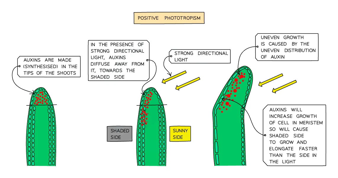
Important: auxin causes existing cells to elongate, not to divide. The number of cells does not change; they just get longer on one side than the other.
Common exam question
Phototropism: the name, the benefit and the experiment
Question: Name a shoot's response to light and the chemical behind it, explain how plants benefit from the responses of stems and roots to light, or describe a simple experiment to show the response (2–4 marks).
Asked in 3 of the 23 papers. The name is positive phototropism; the chemical is auxin (IAA), moving to the shaded side. For the benefit, take stems then roots: stems grow towards light (positively phototropic) for more photosynthesis and to clear competing plants; roots grow away from light (negatively phototropic), into the soil for water and minerals and to anchor the plant. Write stems or shoots: "plants grow towards light" is ignored.
For the experiment, three marks from four points: light one shoot from one side only; keep a comparison shoot in the dark or lit all round; leave both for a stated time with shoot type, temperature and water the same; then record the bending towards the light or measure the angle.
How auxin causes geotropism in a root:
- Auxin in roots collects on the lower side of a horizontal root (due to gravity).
- In roots, high auxin concentration slows cell elongation (unlike in shoots where it speeds it up).
- The cells on the upper side therefore elongate more than the cells on the lower side, and the root bends downwards.
So auxin works in opposite directions in shoots and roots, even though the chemical itself is the same. This nicely explains why shoots curl up towards the sun while roots curl down into the soil.
Practical uses of plant hormones
The discovery of how auxin works has led to several useful agricultural applications:
- Selective weedkillers (herbicides) that contain synthetic auxin-like chemicals make broad-leaved weeds grow uncontrollably until they die. Cereal crops (which have narrow leaves) are mostly unaffected, so farmers can spray a whole field and kill only the weeds.
- Rooting powders for cuttings contain auxin. Dipping the cut end of a stem cutting in rooting powder encourages new roots to grow, which makes propagating plants from cuttings much more reliable.
- Fruit ripening (especially ethene) are used commercially to ripen fruit on demand, so that bananas and tomatoes can be shipped green and ripened just before sale.